
Page 533 - Clinical Small Animal Internal Medicine
P. 533
48 Gastrointestinal Imaging 501
(a) (b)
VetBooks.ir
(c) (d)
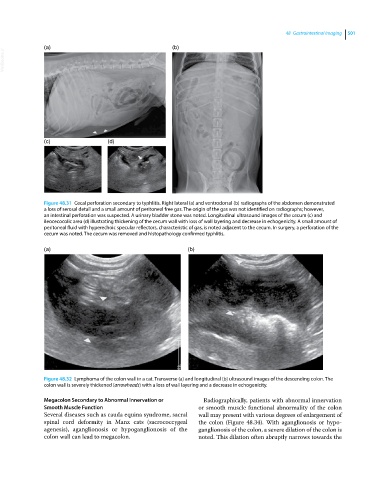
Figure 48.31 Cecal perforation secondary to typhlitis. Right lateral (a) and ventrodorsal (b) radiographs of the abdomen demonstrated
a loss of serosal detail and a small amount of peritoneal free gas. The origin of the gas was not identified on radiographs; however,
an intestinal perforation was suspected. A urinary bladder stone was noted. Longitudinal ultrasound images of the cecum (c) and
ileocecocolic area (d) illustrating thickening of the cecum wall with loss of wall layering and decrease in echogenicity. A small amount of
peritoneal fluid with hyperechoic specular reflectors, characteristic of gas, is noted adjacent to the cecum. In surgery, a perforation of the
cecum was noted. The cecum was removed and histopathology confirmed typhlitis.
(a) (b)
Figure 48.32 Lymphoma of the colon wall in a cat. Transverse (a) and longitudinal (b) ultrasound images of the descending colon. The
colon wall is severely thickened (arrowheads) with a loss of wall layering and a decrease in echogenicity.
Megacolon Secondary to Abnormal Innervation or Radiographically, patients with abnormal innervation
Smooth Muscle Function or smooth muscle functional abnormality of the colon
Several diseases such as cauda equina syndrome, sacral wall may present with various degrees of enlargement of
spinal cord deformity in Manx cats (sacrococcygeal the colon (Figure 48.34). With aganglionosis or hypo
agenesis), aganglionosis or hypoganglionosis of the ganglionosis of the colon, a severe dilation of the colon is
colon wall can lead to megacolon. noted. This dilation often abruptly narrows towards the