
Page 537 - Clinical Small Animal Internal Medicine
P. 537
48 Gastrointestinal Imaging 505
(a) (b) (c)
VetBooks.ir
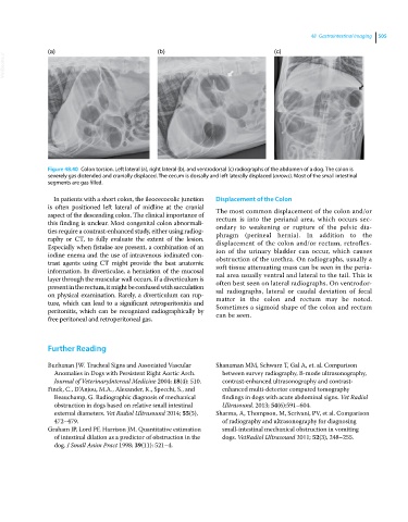
Figure 48.40 Colon torsion. Left lateral (a), right lateral (b), and ventrodorsal (c) radiographs of the abdomen of a dog. The colon is
severely gas distended and cranially displaced. The cecum is dorsally and left laterally displaced (arrows). Most of the small intestinal
segments are gas filled.
In patients with a short colon, the ileocecocolic junction Displacement of the Colon
is often positioned left lateral of midline at the cranial The most common displacement of the colon and/or
aspect of the descending colon. The clinical importance of rectum is into the perianal area, which occurs sec
this finding is unclear. Most congenital colon abnormali ondary to weakening or rupture of the pelvic dia
ties require a contrast‐enhanced study, either using radiog phragm (perineal hernia). In addition to the
raphy or CT, to fully evaluate the extent of the lesion. displacement of the colon and/or rectum, retroflex
Especially when fistulae are present, a combination of an ion of the urinary bladder can occur, which causes
iodine enema and the use of intravenous iodinated con obstruction of the urethra. On radiographs, usually a
trast agents using CT might provide the best anatomic soft tissue attenuating mass can be seen in the peria
information. In diverticulae, a herniation of the mucosal nal area usually ventral and lateral to the tail. This is
layer through the muscular wall occurs. If a diverticulum is often best seen on lateral radiographs. On ventrodor
present in the rectum, it might be confused with sacculation sal radiographs, lateral or caudal deviation of fecal
on physical examination. Rarely, a diverticulum can rup matter in the colon and rectum may be noted.
ture, which can lead to a significant retroperitonitis and Sometimes a sigmoid shape of the colon and rectum
peritonitis, which can be recognized radiographically by can be seen.
free peritoneal and retroperitoneal gas.
Further Reading
Buchanan JW. Tracheal Signs and Associated Vascular Shanaman MM, Schwarz T, Gal A, et. al. Comparison
Anomalies in Dogs with Persistent Right Aortic Arch. between survey radiography, Bmode ultrasonography,
Journal of VeterinaryInternal Medicine 2004; 18(4): 510. contrastenhanced ultrasonography and contrast
Finck, C., D’Anjou, M.A., Alexander, K., Specchi, S., and enhanced multidetector computed tomography
Beauchamp, G. Radiographic diagnosis of mechanical findings in dogs with acute abdominal signs. Vet Radiol
obstruction in dogs based on relative small intestinal Ultrasound. 2013; 54(6):591–604.
external diameters. Vet Radiol Ultrasound 2014; 55(5), Sharma, A, Thompson, M, Scrivani, PV, et al. Comparison
472–479. of radiography and ultrasonography for diagnosing
Graham JP, Lord PF, Harrison JM. Quantitative estimation smallintestinal mechanical obstruction in vomiting
of intestinal dilation as a predictor of obstruction in the dogs. VetRadiol Ultrasound 2011; 52(3), 248–255.
dog. J Small Anim Pract 1998; 39(11): 521–4.