
Page 542 - Clinical Small Animal Internal Medicine
P. 542
510 Section 6 Gastrointestinal Disease
useful lives after burning, and may not survive for the
VetBooks.ir duration necessary to meet legal requirements for record
keeping in some localities.
Given the combination of workflow advantages,
archivability, and integration with other hospital sys-
tems, systems that can achieve digital capture and archiv-
ing are generally the best option currently available, and
will have the greatest future proofing.
Endoscope Handling
General Manipulations
The endoscope is a fragile instrument and needs to be
handled carefully and appropriately to avoid damage
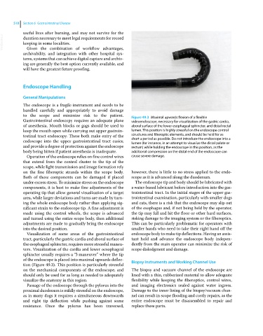
to the scope and minimize risk to the patient. Figure 49.3 Maximal upwards flexion of a flexible
Gastrointestinal endoscopy requires an adequate plane videoendoscope, necessary for visualization of the gastric cardia,
of anesthesia. Mouth blocks or gags should be used to aboral surface of the lower esophageal sphincter, and distal rectal
keep the mouth open while carrying out upper gastroin- lumen. This position is highly stressful on the endoscope control
testinal tract endoscopy. These both make entry of the structures and fiberoptic elements, and should be held for as
endoscope into the upper gastrointestinal tract easier, short a period as possible. Do not introduce the endoscope into a
lumen (for instance, in an attempt to visualize the distal palate or
and provide a degree of protection against the endoscope rectum) while holding the endoscope in this position, as the
body being bitten if patient anesthesia is inadequate. additional compression on the distal end of the endoscope can
Operation of the endoscope relies on fine control wires cause severe damage.
that extend from the control cluster to the tip of the
scope, while light transmission and image formation rely
on the fine fiberoptic strands within the scope body. however, there is little to no stress applied to the endo-
Both of these components can be damaged if placed scope as it is advanced along the duodenum.
under excess stress. To minimize stress on the endoscope The endoscope tip and body should be lubricated with
components, it is best to make fine adjustments of the a water‐based lubricant before introduction into the gas-
operating tip that allow general visualization of a target trointestinal tract. In the initial stages of the upper gas-
area, while larger deviations and turns are made by turn- trointestinal examination, particularly with smaller dogs
ing the whole endoscope body rather than applying sig- and cats, there is a risk that the endoscope may slip out
nificant strain to the endoscope tip. A fine adjustment is of the esophagus and, if not being held by the operator,
made using the control wheels, the scope is advanced the tip may fall and hit the floor or other hard surfaces,
and turned using the entire scope body, then additional risking damage to the imaging system or the fiberoptics.
adjustments are made to gradually bring the endoscope This can be particularly problematic for operators with
into the desired position. smaller hands who need to take their right hand off the
Visualization of some areas of the gastrointestinal endoscope body to make tip deflections. Having an assis-
tract, particularly the gastric cardia and aboral surface of tant hold and advance the endoscope body indepen-
the esophageal sphincter, requires more stressful maneu- dently from the main operator can minimize the risk of
vers. Visualization of the cardia and lower oesophageal scope dislodgment and damage.
sphincter usually requires a “J‐maneuver” where the tip
of the endoscope is placed into maximal upwards deflec- Biopsy Instruments and Working Channel Use
tion (Figure 49.3). This position is particularly stressful
on the mechanical components of the endoscope, and The biopsy and vacuum channel of the endoscope are
should only be used for as long as needed to adequately lined with a thin, rubberized material to allow adequate
visualize the anatomy in this region. flexibility while keeping the fiberoptics, control wires,
Passage of the endoscope through the pylorus into the and imaging electronics sealed against water ingress.
proximal duodenum is mildly stressful on the endoscope, Damage to the inner lining of the biopsy/vacuum chan-
as in many dogs it requires a simultaneous downwards nel can result in scope flooding and costly repairs, as the
and right tip deflection while pushing against some entire endoscope must be disassembled to repair and
resistance. Once the pylorus has been traversed, replace these parts.