
Page 546 - Clinical Small Animal Internal Medicine
P. 546
514 Section 6 Gastrointestinal Disease
(a)
VetBooks.ir
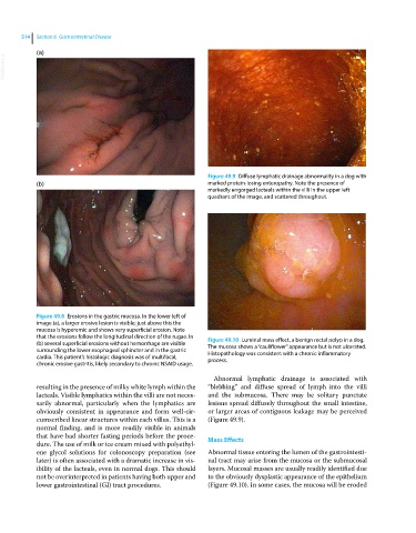
Figure 49.9 Diffuse lymphatic drainage abnormality in a dog with
(b) marked protein‐losing enteropathy. Note the presence of
markedly engorged lacteals within the villi in the upper left
quadrant of the image, and scattered throughout.
Figure 49.8 Erosions in the gastric mucosa. In the lower left of
image (a), a larger erosive lesion is visible; just above this the
mucosa is hyperemic and shows very superficial erosion. Note
that the erosions follow the longitudinal direction of the rugae. In Figure 49.10 Luminal mass effect, a benign rectal polyp in a dog.
(b) several superficial erosions without hemorrhage are visible The mucosa shows a “cauliflower” appearance but is not ulcerated.
surrounding the lower esophageal sphincter and in the gastric Histopathology was consistent with a chronic inflammatory
cardia. This patient’s histologic diagnosis was of multifocal, process.
chronic erosive gastritis, likely secondary to chronic NSAID usage.
Abnormal lymphatic drainage is associated with
resulting in the presence of milky white lymph within the “blebbing” and diffuse spread of lymph into the villi
lacteals. Visible lymphatics within the villi are not neces- and the submucosa. There may be solitary punctate
sarily abnormal, particularly when the lymphatics are lesions spread diffusely throughout the small intestine,
obviously consistent in appearance and form well‐cir- or larger areas of contiguous leakage may be perceived
cumscribed linear structures within each villus. This is a (Figure 49.9).
normal finding, and is more readily visible in animals
that have had shorter fasting periods before the proce- Mass Effects
dure. The use of milk or ice cream mixed with polyethyl-
ene glycol solutions for colonoscopy preparation (see Abnormal tissue entering the lumen of the gastrointesti-
later) is often associated with a dramatic increase in vis- nal tract may arise from the mucosa or the submucosal
ibility of the lacteals, even in normal dogs. This should layers. Mucosal masses are usually readily identified due
not be overinterpreted in patients having both upper and to the obviously dysplastic appearance of the epithelium
lower gastrointestinal (GI) tract procedures. (Figure 49.10). In some cases, the mucosa will be eroded