
Page 548 - Clinical Small Animal Internal Medicine
P. 548
516 Section 6 Gastrointestinal Disease
normal animal; visualization of a strong pulsatile motion In humans, chronic gastroesophageal reflux can lead
VetBooks.ir from these vessels is not necessarily suggestive of a to squamous differentiation of the distal esophageal
mucosa, which is considered a preneoplastic change.
megaesophagus.
The esophageal wall in dogs is usually smooth, or very
chronic gastroesophageal reflux, but the potential for
slightly corrugated, on full insufflation. The cat has a Similar changes are occasionally seen in dogs with
series of mucosal folds in the distal third of the esopha- neoplastic transformation is unknown.
gus that give the normal esophageal lumen a “herring While a relaxed, partially dilated lower esophageal
bone” appearance in this species. sphincter is not necessarily an abnormal finding in dogs
The lower esophageal sphincter is readily visualized in and cats, the prolapse of gastric mucosa into the esopha-
most animals, assuming that there are no impeding for- geal lumen is abnormal. This may occur as a result of
eign bodies. The lower esophageal sphincter is usually severe, chronic reflux esophagitis, hiatal hernias, or be
closed on examination, but a relaxed and open sphincter due to the presence of mass lesions on or just below the
is not necessarily abnormal. Often, a small amount of lower esophageal sphincter (see Gastric Tumors, later).
stable white foam (aerated saliva) is adherent on the dis-
tal esophageal mucosa, which is also normal. Esophageal Foreign Bodies
Esophageal foreign bodies are a significant medical
emergency, and delays in detection and definitive treat-
Potential Abnormalities Observed ment should be avoided as much as possible. Large
in Esophagoscopy Procedures
obstructing foreign bodies, such as bones, swallowed
Esophagitis chew toys, etc., are readily visualized. Common locations
Esophageal inflammation is a common finding in dogs for foreign body entrapment are at the thoracic inlet, cra-
with gastrointestinal disease, particularly if there is gas- nial to the heart base, and cranial to the lower esophageal
troesophageal reflux or chronic vomiting. Changes in sphincter, with the lower esophageal location being the
the esophageal mucosa with esophagitis are highly vari- most common. Sharp, more linear objects such as nee-
able, and often do not correlate closely with the per- dles, fish hooks, etc. may lodge at any point in the esoph-
ceived clinical severity of signs. The most common ageal lumen, but again are most commonly encountered
changes seen with esophagitis are increased granularity at the thoracic inlet and cranial to the lower esophageal
of the esophageal mucosa, hyperemia or erythema, and sphincter. Sharp linear foreign bodies often accumulate
mucosal erosions. Often, these changes are most pro- additional material such as hair, grass, and food frag-
nounced in the distal esophagus, particularly if they are ments that can complicate assessment and removal.
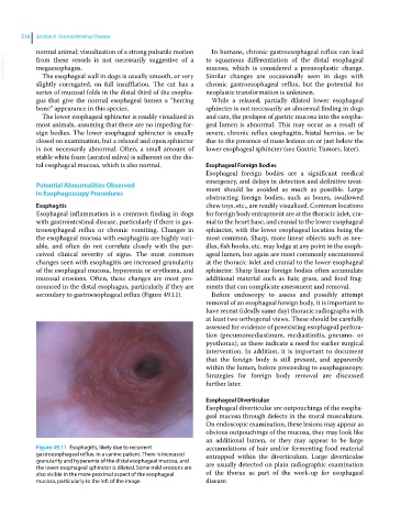
secondary to gastroesophageal reflux (Figure 49.11). Before endoscopy to assess and possibly attempt
removal of an esophageal foreign body, it is important to
have recent (ideally same day) thoracic radiographs with
at least two orthogonal views. These should be carefully
assessed for evidence of preexisting esophageal perfora-
tion (pneumomediastinum, mediastinitis, pneumo‐ or
pyothorax), as these indicate a need for earlier surgical
intervention. In addition, it is important to document
that the foreign body is still present, and apparently
within the lumen, before proceeding to esophagoscopy.
Strategies for foreign body removal are discussed
further later.
Esophageal Diverticulae
Esophageal diverticulae are outpouchings of the esopha-
geal mucosa through defects in the mural musculature.
On endoscopic examination, these lesions may appear as
obvious outpouchings of the mucosa, they may look like
an additional lumen, or they may appear to be large
Figure 49.11 Esophagitis, likely due to recurrent accumulations of hair and/or fermenting food material
gastroesophageal reflux, in a canine patient. There is increased entrapped within the diverticulum. Large diverticulae
granularity and hyperemia of the distal esophageal mucosa, and are usually detected on plain radiographic examination
the lower esophageal sphincter is dilated. Some mild erosions are
also visible in the more proximal aspect of the esophageal of the thorax as part of the work‐up for esophageal
mucosa, particularly to the left of the image. disease.