
Page 545 - Clinical Small Animal Internal Medicine
P. 545
49 Gastrointestinal Endoscopy 513
VetBooks.ir
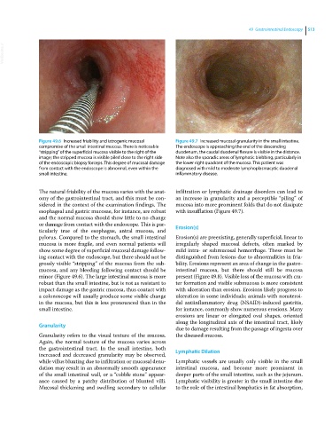
Figure 49.6 Increased friability and iatrogenic mucosal Figure 49.7 Increased mucosal granularity in the small intestine.
compromise of the small intestinal mucosa. There is noticeable The endoscope is approaching the end of the descending
“stripping” of the superficial mucosa visible to the right of the duodenum, the caudal duodenal flexure is visible in the distance.
image; the stripped mucosa is visible piled close to the right side Note also the sporadic areas of lymphatic blebbing, particularly in
of the endoscopic biopsy forceps. This degree of mucosal damage the lower right quadrant of the mucosa. This patient was
from contact with the endoscope is abnormal, even within the diagnosed with mild to moderate lymphoplasmacytic duodenal
small intestine. inflammatory disease.
The natural friability of the mucosa varies with the anat- infiltration or lymphatic drainage disorders can lead to
omy of the gastrointestinal tract, and this must be con- an increase in granularity and a perceptible “piling” of
sidered in the context of the examination findings. The mucosa into more prominent folds that do not dissipate
esophageal and gastric mucosae, for instance, are robust with insufflation (Figure 49.7).
and the normal mucosa should show little to no change
or damage from contact with the endoscope. This is par- Erosion(s)
ticularly true of the esophagus, antral mucosa, and
pylorus. Compared to the stomach, the small intestinal Erosion(s) are preexisting, generally superficial, linear to
mucosa is more fragile, and even normal patients will irregularly shaped mucosal defects, often marked by
show some degree of superficial mucosal damage follow- mild intra‐ or submucosal hemorrhage. These must be
ing contact with the endoscope, but there should not be distinguished from lesions due to abnormalities in fria-
grossly visible “stripping” of the mucosa from the sub- bility. Erosions represent an area of change in the gastro-
mucosa, and any bleeding following contact should be intestinal mucosa, but there should still be mucosa
minor (Figure 49.6). The large intestinal mucosa is more present (Figure 49.8). Visible loss of the mucosa with cra-
robust than the small intestine, but is not as resistant to ter formation and visible submucosa is more consistent
impact damage as the gastric mucosa, thus contact with with ulceration than erosion. Erosions likely progress to
a colonoscope will usually produce some visible change ulceration in some individuals; animals with nonsteroi-
in the mucosa, but this is less pronounced than in the dal antiinflammatory drug (NSAID)‐induced gastritis,
small intestine. for instance, commonly show numerous erosions. Many
erosions are linear or elongated oval shapes, oriented
along the longitudinal axis of the intestinal tract, likely
Granularity
due to damage resulting from the passage of ingesta over
Granularity refers to the visual texture of the mucosa. the diseased mucosa.
Again, the normal texture of the mucosa varies across
the gastrointestinal tract. In the small intestine, both Lymphatic Dilation
increased and decreased granularity may be observed,
while villus blunting due to infiltration or mucosal denu- Lymphatic vessels are usually only visible in the small
dation may result in an abnormally smooth appearance intestinal mucosa, and become more prominent in
of the small intestinal wall, or a “cobble stone” appear- deeper parts of the small intestine, such as the jejunum.
ance caused by a patchy distribution of blunted villi. Lymphatic visibility is greater in the small intestine due
Mucosal thickening and swelling secondary to cellular to the role of the intestinal lymphatics in fat absorption,