
Page 553 - Clinical Small Animal Internal Medicine
P. 553
49 Gastrointestinal Endoscopy 521
VetBooks.ir
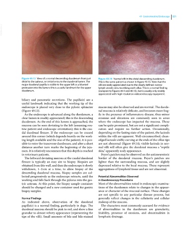
Figure 49.13 View of a normal descending duodenum from just Figure 49.14 Normal villi in the distal descending duodenum.
distal to the pylorus, on initial entry to the duodenal lumen. The This is the same patient as shown in Figure 49.13. Note that the
major duodenal papilla is visible to the upper left as a blunted villi are easily appreciated due to the clearly defined narrow
protrusion into the lumen; this is a useful landmark for the upper lymph vessels circumscribing each villus. This is a normal finding
duodenum. (compare to Figures 49.9 and 49.15), but is usually only readily
appreciated with high‐resolution videoendoscopy equipment.
biliary and pancreatic secretions. The papilla(e) are a
useful landmark indicating that the working tip of the
endoscope is placed very close to the pyloric sphincter mucus may also be observed and are normal. The duode-
(Figure 49.13). nal mucosa is relatively delicate, and becomes more frag-
As the endoscope is advanced along the duodenum, a ile in the presence of inflammatory disease, thus minor
clear lumen is readily appreciated; this is the descending erosions and abrasions are commonly seen in areas
duodenum. As the end of this lumen is approached, the where the endoscope has impacted the mucosa. These
mucosa can be seen deviating to the left (assuming rou- can be quite prominent, but are not a significant compli-
tine patient and endoscope orientation); this is the cau- cation and require no further action. Occasionally,
dal duodenal flexure. If the endoscope can be coaxed depending on the fasting state of the patient, the lacteals
around this corner (which depends heavily on the work- within the villi are apparent. Well‐circumscribed, clean‐
ing length available and the size of the patient), it is pos- edged lacteals visibly curving at the ends of the villus tips
sible to enter the transverse duodenum, and after a short are not abnormal (Figure 49.14); visible lacteals in nor-
distance another turn marks the beginning of the jeju- mal villi will often give the duodenal mucosa a “reptile
num. It is relatively uncommon that this depth is reached skin,” apparently scaly appearance.
in veterinary patients. Peyer’s patches may be observed on the antimesenteric
The leftward deviating mucosa at the caudal duodenal border of the duodenal mucosa. Peyer’s patches are
flexure is typically an easy site to biopsy. Biopsies are lighter than the surrounding mucosa, and are slightly
obtained from this wall, and then the endoscope is slowly depressed relative to the local mucosa. These represent
withdrawn, 1–2 cm at a time, to allow biopsy of the aggregations of lymphoid tissue and are not abnormal.
descending duodenal mucosa. Biopsy samples are col-
lected progressively as the endoscope retracts, until the Potential Abnormalities Observed
working end falls back through the pylorus into the gas- in Duodenoscopy Procedures
tric antrum. At this point, the biopsy sample container Most of the abnormalities noted in endoscopic examina-
should be changed and a new container used for gastric tions of the duodenum relate to changes in the appear-
biopsy samples. ance or character of the mucosal surface. These changes
are not specific to any particular disease process, but
Normal Findings generally reflect changes in the cellularity and cellular
As indicated above, observation of the duodenal makeup of the mucosa.
papilla(e) is a normal finding, particularly in dogs. The The characters most commonly assessed for evidence
duodenal mucosa should be pink to red, and show a fine of abnormalities in the duodenum are granularity,
granular to almost velvety appearance (representing the friability, presence of erosions, and abnormalities in
tips of the villi). Small amounts of bile and bile‐stained lymphatic drainage.