
Page 557 - Clinical Small Animal Internal Medicine
P. 557
49 Gastrointestinal Endoscopy 525
mucosal defects are best assessed by collecting biopsies
VetBooks.ir from the adjacent, more normal‐appearing mucosa
rather than the center of the mass, as biopsies from this
region are usually of poor diagnostic quality due to tissue
necrosis, and the risk of perforation is increased
substantially.
Ileoscopy
Indications
Ileoscopy, while technically more challenging than many
other gastrointestinal endoscopy procedures, is strongly
recommended when endoscopic examination is being
conducted as part of the approach to patients with gas-
trointestinal disease, regardless of whether the primary
Figure 49.16 Colonic view of the normal ileocolic junction in a complaint is predominantly arising from upper or lower
dog. The ileocolic sphincter, visible as a mushroom‐like protrusion gastrointestinal tract signs. It is not unusual to document
into the colonic lumen, occupies the lower half of the image. substantial histological disagreement between duodenal
Above the ileocolic sphincter, the entry to the cecum is visible, and ileal mucosal biopsy specimens, with the ileal
surrounded by a “ridge” of normal colonic mucosa. The anatomy of mucosa often presenting with more severe changes. This
this region, particularly the transition from colonic to cecal lumen,
is highly variable in normal dogs. holds true with both dogs and cats.
Patient Positioning and Preparation
As ileoscopy requires a transcolonic approach, ideally
the patient will be placed in left lateral recumbence. This
is consistent with the positioning for upper gastrointesti-
nal procedures. Ideally, the patient will receive preproc-
edural preparation for colonoscopy. If there is no plan to
fully assess the colonic mucosa and the sole reason for
the procedure is to attempt to biopsy the ileal mucosa, a
more abbreviated preparation protocol can be used,
essentially several warm water enemas administered the
evening before and once on the morning of the proce-
dure (assuming a morning procedure time). This may
provide sufficient space and visualization to allow tra-
versal of the colon to the ileocolic junction, but there is a
greater risk that the lower intensity preparation will
result in inadequate visualization and failure of the
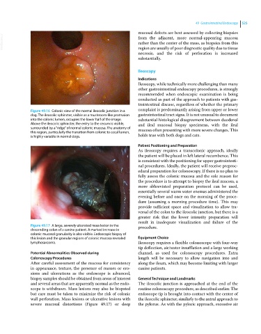
Figure 49.17 A large, severely ulcerated mass lesion in the procedure.
descending colon of a canine patient. A marked increase in
colonic mucosal granularity is also visible. Endoscopic biopsy of
this lesion and the granular regions of colonic mucosa revealed Equipment Choice
lymphosarcoma. Ileoscopy requires a flexible colonoscope with four‐way
tip deflection, air/water insufflation and a large working
Potential Abnormalities Observed during channel, as used for colonoscopy procedures. Extra
Colonoscopy Procedures length will be necessary to allow navigation into and
After careful assessment of the mucosa for consistency along the ileum, which may become limiting with larger
in appearance, texture, the presence of masses or ero- canine patients.
sions and ulcerations as the endoscope is advanced,
biopsy samples should be obtained from areas of interest General Technique and Landmarks
and several areas that are apparently normal as the endo- The ileocolic junction is approached at the end of the
scope is withdrawn. Mass lesions may also be biopsied routine colonoscopy procedure, as described earlier. The
but care must be taken to minimize the risk of colonic endoscope tip is brought into contact with the center of
wall perforation. Mass lesions or ulcerative lesions with the ileocolic sphincter, similarly to the antral approach to
severe mucosal distortions (Figure 49.17) or deep the pylorus. As with the pyloric approach, excessive air