
Page 562 - Clinical Small Animal Internal Medicine
P. 562
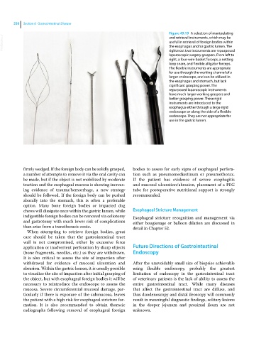
530  Section 6  Gastrointestinal Disease

                                                                               Figure 49.19  A selection of manipulating
  VetBooks.ir                                                                  useful in retrieval of foreign bodies within
                                                                               and retrieval instruments, which may be
                                                                               the esophagus and/or gastric lumen. The
                                                                               rightmost two instruments are repurposed
                                                                               laparoscopic surgery graspers. From left to
                                                                               right, a four‐wire basket forceps, a netting
                                                                               loop snare, and flexible alligator forceps.
                                                                               The flexible instruments are appropriate
                                                                               for use through the working channel of a
                                                                               larger endoscope, and can be utilized in
                                                                               the esophagus and stomach, but lack
                                                                               significant grasping power. The
                                                                               repurposed laparoscopic instruments
                                                                               have much larger working graspers and
                                                                               better grasping power. These rigid
                                                                               instruments are introduced to the
                                                                               esophagus either through a large rigid
                                                                               endoscope or along the side of a flexible
                                                                               endoscope. They are not appropriate for
                                                                               use in the gastric lumen.


            firmly wedged. If the foreign body can be solidly grasped,   bodies to assess for early signs of esophageal perfora-
            a number of attempts to remove it via the oral cavity can   tion such as pneumomediastinum or pneumothorax.
            be made, but if the object is not mobilized by moderate   If  the patient has evidence of severe esophagitis
            traction and the esophageal mucosa is showing increas-  and mucosal ulceration/abrasion, placement of a PEG
            ing evidence of trauma/hemorrhage, a new strategy   tube for postoperative nutritional support is strongly
            should be followed. If the foreign body can be pushed   recommended.
            aborally into the stomach, this is often a preferable
            option. Many bone foreign bodies or impacted dog
            chews will dissipate once within the gastric lumen, while   Esophageal Stricture Management
            indigestible foreign bodies can be removed via celiotomy   Esophageal stricture recognition and management via
            and gastrotomy with much lower risk of complications   either bougienage or balloon dilation are discussed in
            than arise from a transthoracic route.            detail in Chapter 52.
             When attempting to retrieve foreign bodies, great
            care should be taken that the gastrointestinal tract
            wall  is not compromised, either by excessive force
            application or inadvertent perforation by sharp objects     Future Directions of Gastrointestinal
            (bone fragments, needles, etc.) as they are withdrawn.   Endoscopy
            It is also critical to assess the site of impaction after
            withdrawal for evidence of mucosal ulceration and   After the unavoidably small size of biopsies achievable
            abrasion. Within the gastric lumen, it is usually possible   using flexible endoscopy, probably the greatest
            to visualize the site of impaction after initial grasping of     limitation  of endoscopy in the gastrointestinal tract
            the object, but with esophageal foreign bodies it will be   of veterinary patients is the lack of ability to assess the
            necessary to reintroduce the endoscope to assess the   entire gastrointestinal tract. While many diseases
            mucosa. Severe circumferential mucosal damage, par-  that  affect the gastrointestinal tract are diffuse, and
            ticularly if there is exposure of the submucosa, leaves   thus duodenoscopy and distal ileoscopy will commonly
            the patient with a high risk for esophageal stricture for-  result in meaningful diagnostic findings, solitary lesions
            mation. It is also recommended to obtain thoracic   in the deeper jejunum and proximal ileum are not
              radiographs following removal of esophageal foreign   unknown.