
Page 559 - Clinical Small Animal Internal Medicine
P. 559
49 Gastrointestinal Endoscopy 527
Given that each individual sample is very small, and
VetBooks.ir may not be representative of the total of the pathologic
changes present in the patient, it is important to obtain
multiple biopsy samples from each organ sampled.
Assuming adequate biopsy technique and handling, at
least seven samples per site (i.e., seven each from upper
and lower small intestine, stomach) in dogs and 10 in
cats are necessary to achieve acceptable correlation with
full‐thickness biopsy results. If inadequate technique is
used, particularly in cats, there is a very real chance that
samples will have no diagnostic utility.
Biopsy of the gastric mucosa is somewhat more
straightforward than in the small intestine as the stom-
ach is more readily distensible and the greater space
within the organ allows easier manipulation of the endo-
scope to a position perpendicular to the mucosa. The
gastric mucosa is more robust than the small intestinal
mucosa, and good‐quality biopsies are easier to obtain
from most areas of the stomach.
The esophageal mucosa is predominantly squamous
epithelium, and difficult to adequately biopsy unless
severe pathology is present.
Tissues from different sources (small intestine vs large
intestine vs stomach) should be handled and submitted
separately.
Postbiopsy handling of the tissues is also important
for maximum diagnostic utility. Different pathology ser-
vices may have differing requirements for sample sub-
mission but generally speaking, it is best to organize
biopsy samples in a manner that will maintain consist-
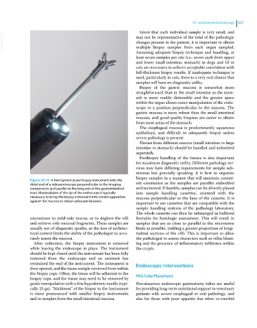
Figure 49.18 A hemispherical jaw biopsy instrument exits the
distal end of a videoendoscope perpendicular to the imaging ent orientation as the samples are paraffin embedded
components and parallel to the long axis of the gastrointestinal and sectioned. If feasible, samples can be directly placed
tract. Manipulation of the tip of the endoscope is typically into sample handling cassettes, oriented with the
necessary to bring the biopsy instrument into correct apposition mucosa perpendicular to the base of the cassette. It is
against the mucosa to obtain adequate biopsies. important to use cassettes that are compatible with the
sample handling systems of the pathology laboratory.
The whole cassette can then be submerged in buffered
uncommon to yield only mucus, or to deglove the villi formalin for histologic assessment. This will result in
and retrieve only mucosal fragments. These samples are samples that are as close to parallel to the microtome
usually not of diagnostic quality, as the loss of architec- blade as possible, yielding a greater proportion of longi-
tural context limits the ability of the pathologist to accu- tudinal sections of the villi. This is important to allow
rately assess the mucosa. the pathologist to assess characters such as villus blunt-
After collection, the biopsy instrument is removed ing and the presence of inflammatory infiltrates within
while leaving the endoscope in place. The instrument the crypts.
should be kept closed until the instrument has been fully
removed from the endoscope and an assistant has
restrained the end of the instrument. The instrument is Endoscopic Interventions
then opened, and the tissue sample retrieved from within
the biopsy cups. Often, the tissue will be adherent to the PEG Tube Placement
biopsy cups, and the tissue may need to be removed by
gentle manipulation with a fine hypodermic needle (typi- Percutaneous endoscopic gastrostomy tubes are useful
cally 25 ga). “Stickiness” of the biopsy to the instrument for providing long‐term nutritional support to veterinary
is more pronounced with smaller biopsy instruments patients with severe esophageal or oral pathology, and
and in samples from the small intestinal mucosa. also for those with poor appetite due other co‐morbid