
Page 554 - Clinical Small Animal Internal Medicine
P. 554
522 Section 6 Gastrointestinal Disease
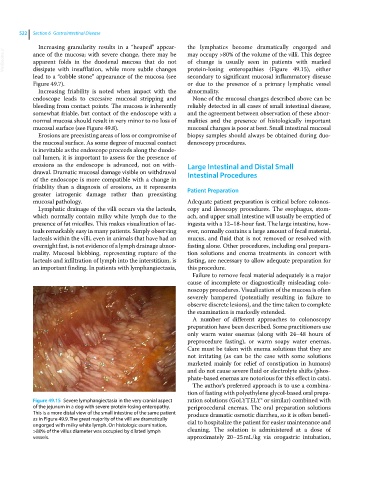
Increasing granularity results in a “heaped” appear- the lymphatics become dramatically engorged and
VetBooks.ir ance of the mucosa; with severe change, there may be may occupy >80% of the volume of the villi. This degree
of change is usually seen in patients with marked
apparent folds in the duodenal mucosa that do not
dissipate with insufflation, while more subtle changes
secondary to significant mucosal inflammatory disease
lead to a “cobble stone” appearance of the mucosa (see protein‐losing enteropathies (Figure 49.15), either
Figure 49.7). or due to the presence of a primary lymphatic vessel
Increasing friability is noted when impact with the abnormality.
endoscope leads to excessive mucosal stripping and None of the mucosal changes described above can be
bleeding from contact points. The mucosa is inherently reliably detected in all cases of small intestinal disease,
somewhat friable, but contact of the endoscope with a and the agreement between observation of these abnor-
normal mucosa should result in very minor to no loss of malities and the presence of histologically important
mucosal surface (see Figure 49.8). mucosal changes is poor at best. Small intestinal mucosal
Erosions are preexisting areas of loss or compromise of biopsy samples should always be obtained during duo-
the mucosal surface. As some degree of mucosal contact denoscopy procedures.
is inevitable as the endoscope proceeds along the duode-
nal lumen, it is important to assess for the presence of
erosions as the endoscope is advanced, not on with- Large Intestinal and Distal Small
drawal. Dramatic mucosal damage visible on withdrawal Intestinal Procedures
of the endoscope is more compatible with a change in
friability than a diagnosis of erosions, as it represents Patient Preparation
greater iatrogenic damage rather than preexisting
mucosal pathology. Adequate patient preparation is critical before colonos-
Lymphatic drainage of the villi occurs via the lacteals, copy and ileoscopy procedures. The esophagus, stom-
which normally contain milky white lymph due to the ach, and upper small intestine will usually be emptied of
presence of fat micelles. This makes visualization of lac- ingesta with a 12–18‐hour fast. The large intestine, how-
teals remarkably easy in many patients. Simply observing ever, normally contains a large amount of fecal material,
lacteals within the villi, even in animals that have had an mucus, and fluid that is not removed or resolved with
overnight fast, is not evidence of a lymph drainage abnor- fasting alone. Other procedures, including oral prepara-
mality. Mucosal blebbing, representing rupture of the tion solutions and enema treatments in concert with
lacteals and infiltration of lymph into the interstitium, is fasting, are necessary to allow adequate preparation for
an important finding. In patients with lymphangiectasia, this procedure.
Failure to remove fecal material adequately is a major
cause of incomplete or diagnostically misleading colo-
noscopy procedures. Visualization of the mucosa is often
severely hampered (potentially resulting in failure to
observe discrete lesions), and the time taken to complete
the examination is markedly extended.
A number of different approaches to colonoscopy
preparation have been described. Some practitioners use
only warm water enemas (along with 24–48 hours of
preprocedure fasting), or warm soapy water enemas.
Care must be taken with enema solutions that they are
not irritating (as can be the case with some solutions
marketed mainly for relief of constipation in humans)
and do not cause severe fluid or electrolyte shifts (phos-
phate‐based enemas are notorious for this effect in cats).
The author’s preferred approach is to use a combina-
tion of fasting with polyethylene glycol‐based oral prepa-
Figure 49.15 Severe lymphangiectasia in the very cranial aspect ration solutions (GoLYTELY® or similar) combined with
of the jejunum in a dog with severe protein‐losing enteropathy. periprocedural enemas. The oral preparation solutions
This is a more distal view of the small intestine of the same patient produce dramatic osmotic diarrhea, so it is often benefi-
as in Figure 49.9. The great majority of the villi are dramatically cial to hospitalize the patient for easier maintenance and
engorged with milky white lymph. On histologic examination,
>80% of the villus diameter was occupied by dilated lymph cleaning. The solution is administered at a dose of
vessels. approximately 20–25 mL/kg via orogastric intubation,