
Page 551 - Clinical Small Animal Internal Medicine
P. 551
49 Gastrointestinal Endoscopy 519
regional prevalence of fungal or oomycete‐induced dis-
VetBooks.ir ease, these infections may also result in intraluminal
granuloma formation. Leiomyoma and leiomyosarcoma
are rare in the gastrointestinal tract but when seen, are
most commonly associated with the lower esophageal
sphincter, and are most frequently identified in older,
female dogs.
As mural mass lesions may primarily affect the deeper
layers of the gastric wall, endoscopic biopsy samples are
often nondiagnostic. Obtaining multiple biopsy “bites”
from the same site, to allow sampling of the submucosa
and muscularis layers, increases the utility of endoscopic
biopsy samples for mural masses.
Gastric Parasites
Occasionally, gastric parasites, such as Physaloptera
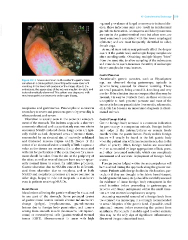
Figure 49.12 Severe ulceration on the wall of the gastric lesser
curvature in a canine patient presenting with severe recurrent spp., are observed during gastroscopy, typically in
vomiting. In the lower left quadrant of the image, close to the patients being assessed for chronic vomiting. These
endoscope, the upper edge of the incisura angularis is visible and are small parasites, being around 3–4 cm long and very
is also dramatically abnormal. This patient was diagnosed with slender. If the clinician does not suspect that they may be
mucinous gastric carcinoma via endoscopic biopsy.
present, it is easy to overlook them. As these worms are
susceptible to both pyrantel pamoate and most of the
macrocylic lactone parasiticides (ivermectin, selamectin,
neoplasms and gastrinomas. Paraneoplastic ulceration etc.), this has become an uncommon diagnosis in client‐
secondary to severe and persistent gastric hyperacidity is owned animals.
often profound and severe.
Ulceration is usually seen in the secretory compart- Gastric Foreign Bodies
ment of the stomach. The incisura angularis is also very Gastric foreign body removal is a common indication
commonly affected, and is a particularly common site to for gastroscopy in companion animals. Foreign bodies
encounter NSAID‐induced ulcers. Large ulcers are typi- may lodge in the antrum/pylorus or remain freely
cally visible as dark, depressed areas of necrotic tissue, mobile within the gastric lumen. Freely mobile foreign
surrounded by an elevated rim of markedly reddened bodies will usually be found in the left gastric body
and thickened mucosa (Figure 49.12). Biopsy of the when the patient is in left lateral recumbence, due to the
center of an ulcerated lesion is usually of little diagnostic effect of gravity. Often, foreign bodies are associated
value as the tissues are necrotic; this is also associated with or surrounded by large aggregations of hair, grass,
with risk for perforation of the ulcer. Biopsies for assess- and other consumed materials, which can complicate
ment should be taken from the rim at the periphery of assessment and accurate deployment of foreign body
the ulcer, as well as several biopsies from nearby appar- snares.
ently normal tissue to screen for infiltrative processes. Foreign bodies lodged within the antrum/pylorus will
Gastric ulceration due to NSAIDs cannot be differenti- be visualized during the approach along the greater cur-
ated from ulceration due to neoplasia, and as both vature. Patients with foreign bodies in this location, par-
NSAID and neoplastic processes are more common in ticularly if they are thought to be fabric based (carpet,
older dogs, biopsy to rule out neoplasia is mandatory bedding material, wool, etc.), should be carefully assessed
even in patients receiving NSAIDs. for evidence of linear foreign body entrapment in the
small intestine before proceeding to gastroscopy, as
Mural Masses patients with linear entrapment within the small intes-
Mass lesions affecting the gastric wall may be visualized tine are best assessed at exploratory surgery.
in any part of the stomach. Some potential causes Assuming successful removal of a foreign body from
of gastric mural lesions include chronic inflammatory the stomach via endoscopy, it is strongly recommended
change (polyps), lymphosarcoma, granulomatous to obtain biopsies of the gastric (and, if possible, small
lesions due to foreign body penetration, and tumors intestinal) mucosa before recovering the patient from
arising from smooth muscle (leiomyoma, leiomyosar- anesthesia. Particularly in middle‐aged to older animals,
coma) or mesenchymal cells (gastrointestinal stromal pica may be the only sign of significant inflammatory
tumor [GIST], fibrosarcomas). In areas with high disease of the gastrointestinal tract.