
Page 534 - Clinical Small Animal Internal Medicine
P. 534
502 Section 6 Gastrointestinal Disease
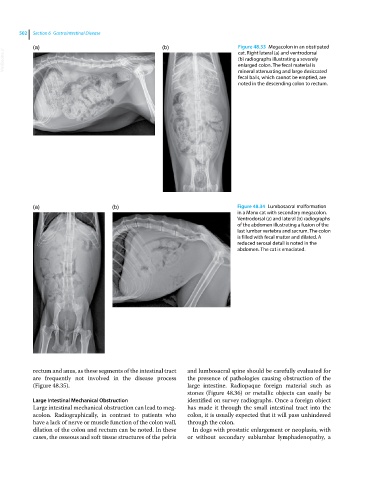
(a) (b) Figure 48.33 Megacolon in an obstipated
VetBooks.ir (b) radiographs illustrating a severely
cat. Right lateral (a) and ventrodorsal
enlarged colon. The fecal material is
mineral attenuating and large desiccated
fecal balls, which cannot be emptied, are
noted in the descending colon to rectum.
(a) (b) Figure 48.34 Lumbosacral malformation
in a Manx cat with secondary megacolon.
Ventrodorsal (a) and lateral (b) radiographs
of the abdomen illustrating a fusion of the
last lumbar vertebra and sacrum. The colon
is filled with fecal matter and dilated. A
reduced serosal detail is noted in the
abdomen. The cat is emaciated.
rectum and anus, as these segments of the intestinal tract and lumbosacral spine should be carefully evaluated for
are frequently not involved in the disease process the presence of pathologies causing obstruction of the
(Figure 48.35). large intestine. Radiopaque foreign material such as
stones (Figure 48.36) or metallic objects can easily be
Large Intestinal Mechanical Obstruction identified on survey radiographs. Once a foreign object
Large intestinal mechanical obstruction can lead to meg has made it through the small intestinal tract into the
acolon. Radiographically, in contrast to patients who colon, it is usually expected that it will pass unhindered
have a lack of nerve or muscle function of the colon wall, through the colon.
dilation of the colon and rectum can be noted. In these In dogs with prostatic enlargement or neoplasia, with
cases, the osseous and soft tissue structures of the pelvis or without secondary sublumbar lymphadenopathy, a