
Page 532 - Clinical Small Animal Internal Medicine
P. 532
500 Section 6 Gastrointestinal Disease
Computed Tomography of the Large Intestine On ultrasound examination, increased thickness of
VetBooks.ir Computed tomography can be an excellent technique for the colon wall with loss of wall layering and decrease of
echogenicity of the colon wall may be noted. Mild
evaluating the large intestine. Similarly to radiography,
contrast is often needed to distend the colon and evalu regional lymph node enlargement may be present.
Typhlitis can be challenging to diagnose clinically as
ate all aspects of the intestinal wall. When the colon is well as via imaging techniques. In mild to moderate
empty and the intestinal walls are collapsed, intestinal typhlitis, no radiographic changes are often noted; in
wall lesions are very difficult or impossible to identify. severe typhlitis on contrast radiographic studies, similar
Additionally, if large amounts of fecal matter are present changes as seen in colitis can be noted. On ultrasound,
in the colon, artifacts created by this might limit visuali thickening of the cecum wall with a decrease of echo
zation of the colon wall. Therefore, similar to radiogra genicity of the wall layers might be noted (Figure 48.31).
phy, optimal patient preparation, including cleansing
enemas, should be performed before a contrast CT study
of the abdomen is performed to evaluate the colon. The Tumors of the Large Intestine
normal anatomy of the colon and regional lymph nodes Tumors of the large intestine account in dogs for more
is easily evaluated using CT and the ability to perform than 60% of the gastrointestinal tumors diagnosed; how
dorsal and sagittal reconstructions of the CT images ever, it is important to remember that the prevalence of
helps to gain a better three‐dimensional understanding gastrointestinal neoplasms in dogs is still only 3–10% of
of normal anatomy or pathologies. The colon wall is thin all tumors diagnosed. Some benign tumors, especially
and measures 1–2 mm in thickness. when polypoid, might not require extensive imaging.
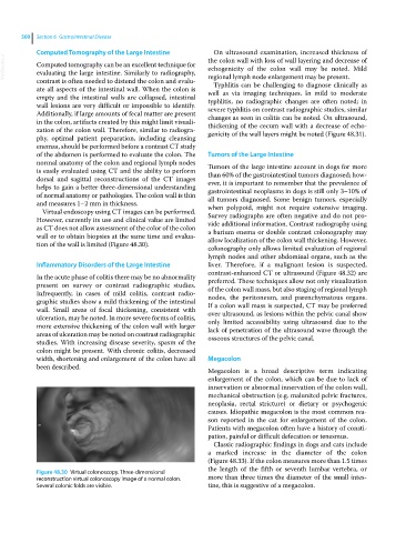
Virtual endoscopy using CT images can be performed.
However, currently its use and clinical value are limited Survey radiographs are often negative and do not pro
vide additional information. Contrast radiography using
as CT does not allow assessment of the color of the colon a barium enema or double contrast colonography may
wall or to obtain biopsies at the same time and evalua allow localization of the colon wall thickening. However,
tion of the wall is limited (Figure 48.30).
colonography only allows limited evaluation of regional
lymph nodes and other abdominal organs, such as the
Inflammatory Disorders of the Large Intestine liver. Therefore, if a malignant lesion is suspected,
contrast‐enhanced CT or ultrasound (Figure 48.32) are
In the acute phase of colitis there may be no abnormality preferred. These techniques allow not only visualization
present on survey or contrast radiographic studies. of the colon wall mass, but also staging of regional lymph
Infrequently, in cases of mild colitis, contrast radio nodes, the peritoneum, and parenchymatous organs.
graphic studies show a mild thickening of the intestinal If a colon wall mass is suspected, CT may be preferred
wall. Small areas of focal thickening, consistent with over ultrasound, as lesions within the pelvic canal show
ulceration, may be noted. In more severe forms of colitis, only limited accessibility using ultrasound due to the
more extensive thickening of the colon wall with larger lack of penetration of the ultrasound wave through the
areas of ulceration may be noted on contrast radiographic osseous structures of the pelvic canal.
studies. With increasing disease severity, spasm of the
colon might be present. With chronic colitis, decreased
width, shortening and enlargement of the colon have all Megacolon
been described.
Megacolon is a broad descriptive term indicating
enlargement of the colon, which can be due to lack of
innervation or abnormal innervation of the colon wall,
mechanical obstruction (e.g. malunited pelvic fractures,
neoplasia, rectal stricture) or dietary or psychogenic
causes. Idiopathic megacolon is the most common rea
son reported in the cat for enlargement of the colon.
Patients with megacolon often have a history of consti
pation, painful or difficult defecation or tenesmus.
Classic radiographic findings in dogs and cats include
a marked increase in the diameter of the colon
(Figure 48.33). If the colon measures more than 1.5 times
the length of the fifth or seventh lumbar vertebra, or
Figure 48.30 Virtual colonoscopy. Three‐dimensional
reconstruction virtual colonoscopy image of a normal colon. more than three times the diameter of the small intes
Several colonic folds are visible. tine, this is suggestive of a megacolon.