
Page 536 - Clinical Small Animal Internal Medicine
P. 536
504 Section 6 Gastrointestinal Disease
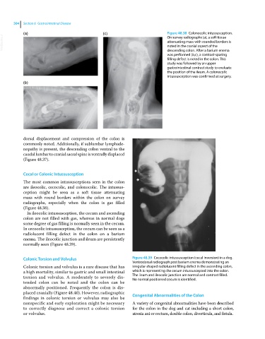
(a) (c) Figure 48.38 Colonocolic intussusception.
VetBooks.ir attenuating mass with rounded borders is
On survey radiographs (a), a soft tissue
noted in the cranial aspect of the
descending colon. After a barium enema
was performed (b,c), a contrast‐sparing
filling defect is noted in the colon. This
study was followed by an upper
gastrointestinal contrast study to evaluate
the position of the ileum. A colonocolic
intussusception was confirmed at surgery.
(b)
dorsal displacement and compression of the colon is
commonly noted. Additionally, if sublumbar lymphade
nopathy is present, the descending colon ventral to the
caudal lumbar to cranial sacral spine is ventrally displaced
(Figure 48.37).
Cecal or Colonic Intussusception
The most common intussusceptions seen in the colon
are ileocolic, cecocolic, and colonocolic. The intussus
ception might be seen as a soft tissue attenuating
mass with round borders within the colon on survey
radiographs, especially when the colon is gas filled
(Figure 48.38).
In ileocolic intussusception, the cecum and ascending
colon are not filled with gas, whereas in normal dogs
some degree of gas filling is normally seen in the cecum.
In cecocolic intussusception, the cecum can be seen as a
radiolucent filling defect in the colon on a barium
enema. The ileocolic junction and ileum are persistently
normally seen (Figure 48.39).
Colonic Torsion and Volvulus Figure 48.39 Cecocolic intussusception (cecal inversion) in a dog.
Ventrodorsal radiograph post barium enema demonstrating an
Colonic torsion and volvulus is a rare disease that has irregular‐shaped radiolucent filling defect in the ascending colon,
a high mortality, similar to gastric and small intestinal which is representing the cecum intussuscepted into the colon.
torsion and volvulus. A moderately to severely dis The ileum and ileocolic junction are normal and contrast filled.
No normal positioned cecum is identified.
tended colon can be noted and the colon can be
abnormally positioned. Frequently the colon is dis
placed cranially (Figure 48.40). However, radiographic Congenital Abnormalities of the Colon
findings in colonic torsion or volvulus may also be
nonspecific and early exploration might be necessary A variety of congenital abnormalities have been described
to correctly diagnose and correct a colonic torsion for the colon in the dog and cat including a short colon,
or volvulus. atresia ani or rectum, double colon, diverticula, and fistula.