
Page 517 - Clinical Small Animal Internal Medicine
P. 517
48 Gastrointestinal Imaging 485
If gastric dilation volvulus is present, no further imaging gastropathy may reveal delayed gastric emptying, focal
VetBooks.ir is necessary and emergency procedures should be per intraluminal contrast sparing areas in the pyloric antrum
with incomplete filling and blunting of the pyloric canal. If
formed to decompress the stomach.
contrast medium remains in the stomach 3–4 hours after
administration, this usually indicates pyloric obstructive
Chronic Gastric Outlet/Pyloric Obstruction
disease. Intraluminal filling defects in the pyloric antrum
Chronic gastric outlet (pyloric) obstruction occurs rela are often caused by thickened mucosal fold(s) extending
tively frequently secondary to either congenital or acquired into the lumen. The shape and radiographic appearance of
pyloric stenosis or abnormal function of the pylorus. the contrast agent filling of the pylorus have been described
Pyloric obstruction is more frequently encountered in with various signs including the “beak,” “tit,” and “string”
dogs than in cats. Radiographic recognition of gastric out signs. Lack of filling or only partial filling of the pyloric
let obstruction often depends on the degree of distension canal has been described as the “beak” sign, where only a
and content of the stomach, and duration of the disease. small amount of contrast agent extends in the form of a
Furthermore, positioning of the patient during radiogra beak into the pyloric canal. The “tit” sign has been used
phy and frequency of vomiting affect the radiographic when a small outpouching remains at the lesser curvature
appearance of pyloric obstruction. Most frequently, the secondary to peristaltic waves. The “string” sign has been
stomach is distended with gas and fluid, which can easily be used when contrast agent extends through the pyloric
noted on radiographs (Figure 48.15). It is important to canal but is concentrically narrowed secondary to circum
remember that not all patients with a gastric dilation have ferential thickening of the pylorus wall.
a mechanical obstruction of the pylorus. On an ultrasound exam, mucosal or muscular thicken
Gastric dilation may also occur secondary to mechan ing of the pylorus wall may be noted and may help to
ical obstruction of the duodenum or with concurrent differentiate between various etiologies of chronic outlet
pancreatitis. Left recumbent lateral views can help to obstruction. In dogs with chronic pyloric hypertrophy,
decide if a duodenal obstructive lesion is present, as gas the hypertrophic muscularis may appear as a thick,
in the duodenum may be able to outline the cause of the hypoechoic layer (Figures 48.15 and 48.16). Sonogra
obstruction. Additionally, in chronic obstructive gas phically, the measured thickness of the muscularis may
tropathy a “gravel sign” indicating retention of small help to categorize disease severity. The muscularis thick
mineral attenuating material particles in the stomach ness in the pylorus is greater than 3 mm in mild to
can sometimes be seen. moderate hypertrophy and greater than 8 mm in severe
Contrast studies of the stomach or an ultrasound chronic pyloric stenosis in dogs. Forceful, but ineffective,
examination are often needed to establish the cause and peristaltic contractions that fail to propel contents into
site of outflow obstruction of the stomach. Contrast radiog the duodenum may be seen in some patients with gastric
raphy of the stomach in cases of chronic obstructive outflow obstruction.
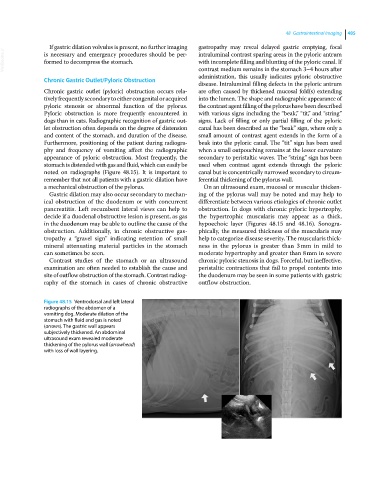
Figure 48.15 Ventrodorsal and left lateral
radiographs of the abdomen of a
vomiting dog. Moderate dilation of the
stomach with fluid and gas is noted
(arrows). The gastric wall appears
subjectively thickened. An abdominal
ultrasound exam revealed moderate
thickening of the pylorus wall (arrowhead)
with loss of wall layering.