
Page 796 - Withrow and MacEwen's Small Animal Clinical Oncology, 6th Edition
P. 796
774 PART IV Specific Malignancies in the Small Animal Patient
The heart is the second most common primary site for canine
HSA and is the most common cardiac neoplasm in dogs. HSA
most commonly originates from the right atrium or auricle;
VetBooks.ir however, other cardiac sites have been reported. 15,35–37 Although
previously thought to be a rather frequent occurrence based on
necropsy studies, one study showed the presence of concurrent
splenic and cardiac HSA to be uncommon (8.7%). 53
Although typically aggressive, the biologic behavior of HSA
can vary depending on primary tumor location, as certain primary
HSA sites, specifically the skin, can be associated with a less aggres-
sive disease course. The more common visceral forms are char-
14
acterized by local infiltration and metastatic dissemination early
in the course of disease. Metastasis occurs either hematogenously
or via intracavitary implantation after tumor rupture. Metastasis
can occur at any site; however, the liver, omentum, peritoneum,
and lungs are the most frequent sites of dissemination. 2–4 In dogs,
HSA is the most common tumor to metastasize to the brain. 54
In the cat, cutaneous and visceral (e.g., spleen, liver, intes-
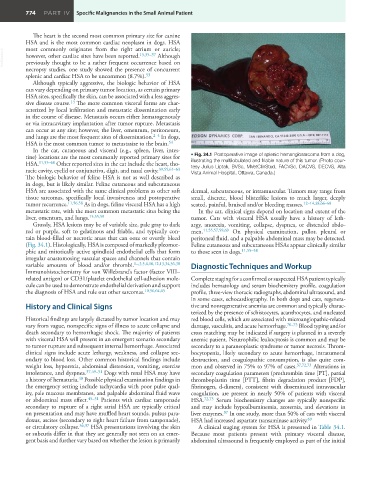
tine) locations are the most commonly reported primary sites for • Fig. 34.1 Postoperative image of splenic hemangiosarcoma from a dog,
HSA. 11,55–60 Other reported sites in the cat include the heart, tho- illustrating the multilobulated and friable nature of this tumor. (Photo cour-
racic cavity, eyelid or conjunctiva, digit, and nasal cavity. 39,55,61–63 tesy Julius Liptak, BVSc, MVetClinStud, FACVSc, DACVS, DECVS, Alta
Vista Animal Hospital, Ottawa, Canada.)
The biologic behavior of feline HSA is not as well described as
in dogs, but is likely similar. Feline cutaneous and subcutaneous
HSA are associated with the same clinical problems as other soft dermal, subcutaneous, or intramuscular. Tumors may range from
tissue sarcomas, specifically local invasiveness and postoperative small, discrete, blood blisterlike lesions to much larger, deeply
tumor recurrence. 11,56,58 As in dogs, feline visceral HSA has a high seated, painful, bruised and/or bleeding masses. 12–14,38,66–68
metastatic rate, with the most common metastatic sites being the In the cat, clinical signs depend on location and extent of the
liver, omentum, and lungs. 15,55,59 tumor. Cats with visceral HSA usually have a history of leth-
Grossly, HSA lesions may be of variable size, pale gray to dark argy, anorexia, vomiting, collapse, dyspnea, or distended abdo-
red or purple, soft to gelatinous and friable, and typically con- men. 11,55,57,59,69 On physical examination, pallor, pleural or
tain blood-filled or necrotic areas that can ooze or overtly bleed peritoneal fluid, and a palpable abdominal mass may be detected.
(Fig. 34.1). Histologically, HSA is composed of markedly pleomor- Feline cutaneous and subcutaneous HSAs appear clinically similar
phic and mitotically active spindloid endothelial cells that form to those seen in dogs. 11,55–58
irregular anastomosing vascular spaces and channels that contain
variable amounts of blood and/or thrombi. 1–,3,5,6,10,12,13,34,36,38 Diagnostic Techniques and Workup
Immunohistochemistry for von Willebrand’s factor (factor VIII–
related antigen) or CD31/platelet endothelial cell-adhesion mole- Complete staging for a confirmed or suspected HSA patient typically
cule can be used to demonstrate endothelial derivation and support includes hematology and serum biochemistry profile, coagulation
the diagnosis of HSA and rule out other sarcomas. 10,56,64,65 profile, three-view thoracic radiographs, abdominal ultrasound, and
in some cases, echocardiography. In both dogs and cats, regenera-
History and Clinical Signs tive and nonregenerative anemias are common and typically charac-
terized by the presence of schistocytes, acanthocytes, and nucleated
Historical findings are largely dictated by tumor location and may red blood cells, which are associated with microangiopathic-related
vary from vague, nonspecific signs of illness to acute collapse and damage, vasculitis, and acute hemorrhage. 70–73 Blood typing and/or
death secondary to hemorrhagic shock. The majority of patients cross matching may be indicated if surgery is planned in a severely
with visceral HSA will present in an emergent scenario secondary anemic patient. Neutrophilic leukocytosis is common and may be
to tumor rupture and subsequent internal hemorrhage. Associated secondary to a paraneoplastic syndrome or tumor necrosis. Throm-
clinical signs include acute lethargy, weakness, and collapse sec- bocytopenia, likely secondary to acute hemorrhage, intratumoral
ondary to blood loss. Other common historical findings include destruction, and coagulopathic consumption, is also quite com-
weight loss, hyporexia, abdominal distension, vomiting, exercise mon and observed in 75% to 97% of cases. 37,72,73 Alterations in
intolerance, and dyspnea. 37,49–51 Dogs with renal HSA may have secondary coagulation parameters (prothrombin time [PT], partial
40
a history of hematuria. Possible physical examination findings in thromboplastin time [PTT], fibrin degradation product [FDP],
the emergency setting include tachycardia with poor pulse qual- fibrinogen, d-dimers), consistent with disseminated intravascular
ity, pale mucous membranes, and palpable abdominal fluid wave coagulation, are present in nearly 50% of patients with visceral
or abdominal mass effect. 49–51 Patients with cardiac tamponade HSA. 72,73 Serum biochemistry changes are typically nonspecific
secondary to rupture of a right atrial HSA are typically critical and may include hypoalbuminemia, azotemia, and elevations in
37
on presentation and may have muffled heart sounds, pulsus para- liver enzymes. In one study, more than 50% of cats with visceral
doxus, ascites (secondary to right heart failure from tamponade), HSA had increased aspartate transaminase activity. 69
or circulatory collapse. 36,37 HSA presentations involving the skin A clinical staging system for HSA is presented in Table 34.1.
or subcutis differ in that they are generally not seen on an emer- Because most patients present with primary visceral disease,
gent basis and further vary based on whether the lesion is primarily abdominal ultrasound is frequently employed as part of the initial