
Page 797 - Withrow and MacEwen's Small Animal Clinical Oncology, 6th Edition
P. 797
CHAPTER 34 Miscellaneous Tumors 775
TABLE 34.1 Clinical Staging System for Canine
Hemangiosarcoma
VetBooks.ir Primary Tumor (T)
T0
No evidence of tumor
T1 Tumor less than 5 cm diameter and confined to primary tissues
T2 Tumor 5 cm or greater or ruptured, invading subcutaneous
tissues
T3 Tumor invading adjacent structures, including muscle
Regional Lymph Nodes (N)
N0 No regional lymph node involvement
N1 Regional lymph node involvement
N2 Distant lymph node involvement
Distant Metastasis (M)
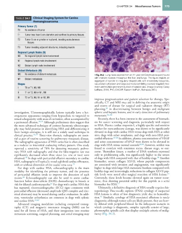
M0 No evidence of distant metastasis • Fig. 34.2 Lung mass aspirate from an 11-year-old Catahoula leopard hound
M1 Distant metastasis with multiple masses throughout the liver and lungs. The figure depicts an
aggregate of spindle to irregularly shaped cells with moderately basophilic,
Stages vacuolated cytoplasm and large oval nuclei exhibiting coarsely stippled chro-
I T0 or T1, N0, M0 matin and multiple prominent nucleoli of variable size. (Image courtesy Casey
LeBlanc, DVM, PhD, DACVP, Eastern VetPath, Bethesda, MD.)
II T1 or T2, N0 or N1, M0
III T2 or T3, N0, N1 or N2, M1
improve prognostication and patient selection for therapy. Spe-
cifically, CT and MRI may aid in defining the anatomic origin
and extent of disease for surgical and radiation therapy (RT)
83
planning, in discriminating between benign and malignant
investigation. Ultrasonographically, lesions typically have a het- splenic and hepatic lesions, and in early detection of pulmonary
erogeneous appearance ranging from hypoechoic to targetoid to metastasis. 84–86
mixed echogenicity with areas of cavitation, often accompanied by Recently, there has been interest in the assessment of biomark-
a peritoneal effusion. 74,75 Although preliminary data suggest that ers for cancer screening and diagnosis, particularly with respect
the more advanced technique of contrast harmonic ultrasonogra- to HSA. Plasma cardiac troponin I, a highly specific and sensitive
phy may hold promise in identifying HSA and differentiating it marker for myocardiocyte damage, was shown to be significantly
from benign etiologies, it is still not a widely used technique in elevated in dogs with cardiac HSA versus dogs with HSA at other
clinical practice. 76,77 Three-view thoracic radiographs are essen- sites, dogs with other neoplasms, and dogs with non-HSA peri-
tial as part of routine screening for pulmonary metastatic disease. cardial effusions. 87,88 In addition, plasma concentrations of VEGF
The radiographic appearance of HSA varies but is often described and urine concentrations of bFGF were shown to be elevated in
as a nodular to interstitial coalescing miliary pattern. One study dogs with HSA versus normal controls 32,89 ; however, neither was
reported a sensitivity of 78% for detecting metastatic pulmo- found to correlate with remission status, disease stage, or out-
nary HSA with radiography and that the false-negative rate was come. Thymidine kinase, a marker of DNA synthesis expressed
significantly decreased when three views (vs. one or two) were only in proliferating cells, was significantly higher in the serum
78
90
obtained. In dogs with pericardial effusion secondary to cardiac of dogs with HSA compared with that of healthy dogs. Another
HSA, radiographs will typically reveal a globoid cardiac silhouette, biomarker, serum collagen XXVII, whose peptide components
with or without distension of the caudal vena cava. 79 are associated with invasion and angiogenesis, was significantly
For dogs with cardiac HSA, echocardiography is the main higher in dogs with large HSA metastatic burdens compared with
modality for identifying the primary tumor, and the presence healthy dogs and interestingly, reductions in collagen XXVII pep-
91
of pericardial effusion tends to improve the detection of such tide levels were noted after surgical resection of HSA lesions.
80
masses. Echocardiography can also be used to assess cardiac Conversely, these levels became elevated again on tumor recur-
function before doxorubicin (DOX) chemotherapy in breeds at rence, thus showing potential utility for this peptide as a serial
81
risk for dilated cardiomyopathy. For dogs with cardiac HSA that biomarker for HSA. 91
has ruptured, electrocardiographic (ECG) signs consistent with Ultimately, a definitive diagnosis of HSA usually requires his-
pericardial effusion (decreased amplitude QRS complex and elec- topathology. Fine-needle aspirate (FNA) cytology of suspected
trical alternans) may be noted during cardiac evaluation. In addi- HSA lesions is often of low diagnostic yield due to hemodilu-
92
tion, ventricular arrhythmias are common in dogs with splenic tion. Similarly, cytology of HSA-associated effusions is rarely
and cardiac HSA. 35,82 diagnostic; although tumor cells are likely present, they are heav-
Advanced imaging modalities including computed tomog- ily diluted with peripheral blood. In the infrequent scenario in
raphy (CT) and magnetic resonance imaging (MRI) can be which cytology is diagnostic, samples typically consist of large,
used for all forms of HSA, and their integration into routine pleomorphic spindle cells that display multiple criteria of malig-
metastasis screening, surgical planning, and serial restaging may nancy (Fig. 34.2). 