
Page 320 - Small Animal Clinical Nutrition 5th Edition
P. 320
Feeding Working and Sporting Dogs 327
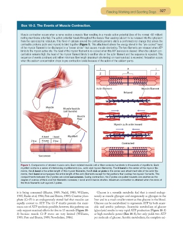
VetBooks.ir Box 18-2. The Events of Muscle Contraction.
Muscle contraction occurs when a nerve excites a muscle fiber resulting in a muscle action potential (loss of the normal -90 millivolt
resting membrane potential). The action potential travels throughout the muscle fiber causing calcium to be released into the cytoplasm
from the sarcoplasmic reticulum. This flood of calcium around the contractile proteins elicits a conformational change that allows the
contractile proteins (actin and myosin) to bind together (Figure 1). This attachment allows the energy stored in the “pre-cocked” head
of the myosin filament to be discharged in a “power stroke” that causes muscle shortening. The two filaments are released when ATP
binds to the myosin active site. The head of the myosin filament is re-cocked when this ATP molecule is cleaved. When the calcium con-
centration remains high, the head of the myosin filament binds to another site on the actin filament and the sequence is repeated. This
sequence of events continues until either minimum fiber length (maximum shortening) or maximum load is exceeded. Relaxation occurs
when the calcium concentration drops to pre-contraction levels because of the action of the calcium pump.
Figure 1. Components of striated muscle cells. Each striated muscle cell or fiber contains hundreds to thousands of myofibrils. Each
myofibril contains a series of interlocking myofilaments (i.e., actin and myosin filaments). The H band is the center of the myosin fila-
ments, the A band is the entire length of the myosin filaments, the Z disk or plate is the center and attachment site of the actin fila-
ments, the I band encompasses the entire length of the actin filaments except for the portions that overlap the myosin filaments. The
compartments between the Z plates are called sarcomeres. During contraction, the Z plates are pulled towards one another as the
degree of overlap of thick and thin filaments increases; I band and H bands shorten. Maximum contraction is attained when the ends of
the thick filaments butt against Z plates.
it is being consumed (Blaxter, 1989; Nadel, 1985; Williams, Glucose is a versatile metabolic fuel that is stored endoge-
1985; Rusko et al, 1986; Pate and Brunn, 1989). Creatine phos- nously as muscle glycogen and exogenously as glycogen in the
phate (Cr-P) is an endogenously stored fuel that muscles can liver and to a much smaller extent as free glucose in the blood.
rapidly convert to ATP. The Cr-P shuttle permits the maxi- Glucose can be metabolized to regenerate ATP by both anaer-
mum rate of ATP synthesis possible; however, this pathway can obic and aerobic pathways. Anaerobic metabolism of glucose
only support maximal efforts for five to 15 seconds (Figure 18- (glycolysis) results in very rapid ATP production (Figure 18-3)
3) because muscle Cr-P stores are very limited (Williams, or high metabolic power (Box 18-3), but only yields two ATP
1985; Pate and Brunn, 1989; Newsholme, 1986). per molecule of glucose. Aerobic metabolism, the complete oxi-