
Page 875 - Small Animal Clinical Nutrition 5th Edition
P. 875
906 Small Animal Clinical Nutrition
VetBooks.ir
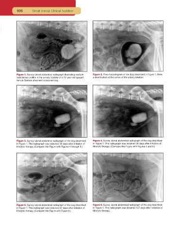
Figure 1. Survey lateral abdominal radiograph illustrating multiple Figure 2. Pneumocystogram of the dog described in Figure 1. Note
radiodense uroliths in the urinary bladder of a 12-year-old spayed a diverticulum at the vertex of the urinary bladder.
female German shepherd crossbred dog.
Figure 3. Survey lateral abdominal radiograph of the dog described Figure 4. Survey lateral abdominal radiograph of the dog described
in Figure 1. This radiograph was obtained 30 days after initiation of in Figure 1. This radiograph was obtained 58 days after initiation of
litholytic therapy. (Compare this Figure with Figures 4 through 6.) litholytic therapy. (Compare this Figure with Figures 5 and 6.)
Figure 5. Survey lateral abdominal radiograph of the dog described Figure 6. Survey lateral abdominal radiograph of the dog described
in Figure 1. This radiograph was obtained 97 days after initiation of in Figure 1. This radiograph was obtained 127 days after initiation of
litholytic therapy. (Compare this Figure with Figure 6.) litholytic therapy.