Page 562 - Feline diagnostic imaging
P. 562
31.3 SAc Diiss sSopeiDe SAects Sody epp 575
(a) (b)
(c)
(e)
(d)
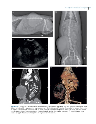
Figure 31.27 A 6-year-old DSH presented for a rapidly growing mass over the right caudal body wall. Abdominal radiographic lateral
(a) and ventrodorsal (b) images show the large mass overlying the right body wall. Abdominal ultrasound of the mass (c) shows a rim
of soft tissue surrounding an anechoic fluid-filled center. Computed tomographic postcontrast reformatted dorsal images (d) show a
rim of contrast enhancement surrounding soft tissue and assist in surgical planning. The reformatted CT image (e) highlights the
vascular supply to the tumor. The histopathologic diagnosis was fibrosarcoma.

