
Page 563 - Feline diagnostic imaging
P. 563
(a) (b)
(c)
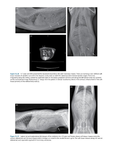
Figure 31.28 A 7-year-old DSH presented for increased respiratory rate and mammary masses. There are numerous well-defined soft
tissue nodules of variable size within the thoracic cavity seen on both the lateral (a) and ventrodorsal (b) images. There was
irregularity along the ventral abdominal wall best seen on the lateral projection (arrows) and along the left lateral body wall (arrows)
on the ventrodorsal image. Postcontrast CT image with the patient in dorsal recumbency shows mild contrast enhancement of the soft
tissue (arrows) on the ventral body wall (c).
(b)
(a)
Figure 31.29 Lateral (a) and ventrodorsal (b) images of the abdomen in a 15-year-old Persian shows soft tissue masses along the
ventral abdominal wall (arrows). Degenerative changes are noted in the caudal thoracic spine. The soft tissue masses along the ventral
abdominal wall represent regrowth of mammary carcinoma.