
Page 1201 - Small Animal Clinical Nutrition 5th Edition
P. 1201
Nutrition of Reptiles 1251
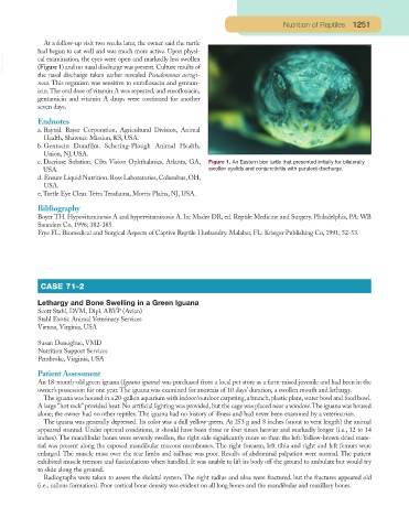
At a follow-up visit two weeks later, the owner said the turtle
had begun to eat well and was much more active. Upon physi-
VetBooks.ir cal examination, the eyes were open and markedly less swollen
(Figure 1) and no nasal discharge was present. Culture results of
the nasal discharge taken earlier revealed Pseudomonas aerugi-
nosa. This organism was sensitive to enrofloxacin and gentam-
icin.The oral dose of vitamin A was repeated, and enrofloxacin,
gentamicin and vitamin A drops were continued for another
seven days.
Endnotes
a. Baytril. Bayer Corporation, Agricultural Division, Animal
Health, Shawnee Mission, KS, USA.
b. Gentocin Durafilm. Schering-Plough Animal Health,
Union, NJ, USA.
c. Dacriose Solution. Ciba Vision Ophthalmics, Atlanta, GA, Figure 1. An Eastern box turtle that presented initially for bilaterally
USA. swollen eyelids and conjunctivitis with purulent discharge.
d. Ensure Liquid Nutrition. Ross Laboratories, Columbus, OH,
USA.
e. Turtle Eye Clear. Tetra Terafauna, Morris Plains, NJ, USA.
Bibliography
Boyer TH. Hypovitaminosis A and hypervitaminosis A. In: Mader DR, ed. Reptile Medicine and Surgery. Philadelphia, PA: WB
Saunders Co, 1996; 382-385.
Frye FL. Biomedical and Surgical Aspects of Captive Reptile Husbandry. Malabar, FL: Krieger Publishing Co, 1991; 52-53.
CASE 71-2
Lethargy and Bone Swelling in a Green Iguana
Scott Stahl, DVM, Dipl. ABVP (Avian)
Stahl Exotic Animal Veterinary Services
Vienna, Virginia, USA
Susan Donoghue, VMD
Nutrition Support Services
Pembroke, Virginia, USA
Patient Assessment
An 18-month-old green iguana (Iguana iguana) was purchased from a local pet store as a farm-raised juvenile and had been in the
owner’s possession for one year. The iguana was examined for anorexia of 10 days’ duration, a swollen mouth and lethargy.
The iguana was housed in a 20-gallon aquarium with indoor/outdoor carpeting,a branch,plastic plant,water bowl and food bowl.
A large “hot rock” provided heat. No artificial lighting was provided, but the cage was placed near a window.The iguana was housed
alone; the owner had no other reptiles. The iguana had no history of illness and had never been examined by a veterinarian.
The iguana was generally depressed. Its color was a dull yellow-green. At 253 g and 8 inches (snout to vent length) the animal
appeared stunted. Under optimal conditions, it should have been three or four times heavier and markedly longer (i.e., 12 to 14
inches). The mandibular bones were severely swollen, the right side significantly more so than the left. Yellow-brown dried mate-
rial was present along the exposed mandibular mucous membranes. The right forearm, left tibia and right and left femurs were
enlarged. The muscle mass over the rear limbs and tailbase was poor. Results of abdominal palpation were normal. The patient
exhibited muscle tremors and fasciculations when handled. It was unable to lift its body off the ground to ambulate but would try
to slide along the ground.
Radiographs were taken to assess the skeletal system. The right radius and ulna were fractured, but the fractures appeared old
(i.e., callous formation). Poor cortical bone density was evident on all long bones and the mandibular and maxillary bones.