
Page 489 - Small Animal Clinical Nutrition 5th Edition
P. 489
Obesity 505
VetBooks.ir Box 27-2. Other Diagnostic Procedures.
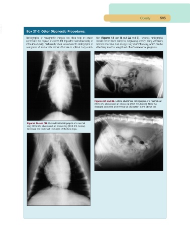
Radiographic or sonographic images can often help an owner tion (Figures 1A and B and 2A and B). However, radiographs
appreciate the degree of excess fat deposited subcutaneously or should not be taken solely for diagnosing obesity. Many veterinary
intra-abdominally, particularly when viewed next to radiographs or schools now have dual energy x-ray absorptiometry, which can be
sonograms of similar size animals that are in optimal body condi- effectively used for weight-reduction/maintenance programs.
Figures 2A and 2B. Lateral abdominal radiographs of a normal cat
(BCS 3/5, above) and an obese cat (BCS 5/5, below). Note the
enlarged abdomen and ventral fat deposition in the obese cat.
Figures 1A and 1B. Ventrodorsal radiographs of a normal
dog (BCS 3/5, above) and an obese dog (BCS 5/5, below).
Compare the body wall thickness of the two dogs.