
Page 637 - Small Animal Clinical Nutrition 5th Edition
P. 637
Skin and Hair Disorders 659
Assess the Food and Feeding Method
VetBooks.ir For the three years before presentation, the dog had eaten a com-
mercial specialty brand dry dog food supplemented with table
foods.
Therapy Including Feeding Plan
The tentative diagnosis was vitamin A-responsive dermatosis
with superficial pyoderma and yeast otitis.Treatment was initiat-
ed with 10,000 IU of vitamin A orally along with the dog’s orig-
inal food.The patient was also given an appropriate antibiotic for
the pyoderma and a topical antifungal for the yeast otitis.
Questions
1. Why is vitamin A essential for normal epidermal function?
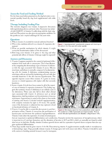
2. Why is this condition referred to as vitamin A-responsive der- Figure 1. Hyperpigmented, hyperkeratotic plaques with fronding on
matosis? the ventrum of a four-year-old cocker spaniel.
3. What are possible mechanisms by which vitamin A might
correct the keratinization defect of this dermatosis?
4. How long must vitamin A be given to this dog and what
potential side effects of vitamin A therapy might be expected?
Answers and Discussion
1. Vitamin A appears essential in the control of epidermal differ-
entiation from basal cells to corneocytes. This is best illustrat-
ed by comparing the dermatologic signs of vitamin A deficien-
cy with the signs associated with vitamin A excess. Mucous
membrane epithelium is normally composed of nonkeratiniz-
ing cells. In vitamin A deficiency, nonkeratinizing mucous
membrane cells are replaced by keratinizing cells and cells that
normally keratinize in the skin become hyperkeratotic. The
opposite response occurs when vitamin A is given in excess;
mucous or ciliated squamous cells replace cells that normally
keratinize.
2. Serum vitamin A levels have been normal in all of the report-
ed cases of vitamin A-responsive dermatosis.This finding sug-
gests that systemic vitamin A deficiency is an unlikely cause of
the dermatosis. These cases also fail to show other clinical
signs associated with vitamin A deficiency such as retinal
degeneration, hind leg weakness and keratinization of mucous
membranes. Improvement is noted within three to four weeks
of starting oral vitamin A alcohol (retinol) supplementation,
with complete remission by eight to 10 weeks. The specific
cause of the dermatosis is unknown but may represent a local
or functional deficiency of vitamin A.
3. Vitamin A may be able to correct this dermatosis via anti-ker-
Figure 2. Skin biopsy specimen from a seborrheic cocker spaniel.
atinization effects. Vitamin A normalizes the proliferation of The epidermis is mildly hyperplastic and hyperkeratotic. There is
keratinocytes and decreases the epidermal hyperproliferation. severe follicular hyperkeratosis and dilatation. (Magnification 10X.)
Vitamin A also alters the expression of certain structural genes
that are important in epidermal differentiation and cornification. Examples include the suppression of transglutaminase, which
is important in cell envelope formation, and the alteration of keratins to K19 and K13, which are normally not found in adult
skin but are in fetal skin. Finally, vitamin A induces growth factors and the expression of growth factor receptors that also sup-
press epidermal differentiation.
4. These dogs usually must be given vitamin A for life. Discontinuing vitamin A supplementation usually results in recrudescence
of dermatologic signs. Dogs generally tolerate vitamin A therapy quite well with minor side effects. Vitamin A should be used
with caution in breeding animals because it may be teratogenic.