
Page 641 - Small Animal Clinical Nutrition 5th Edition
P. 641
Skin and Hair Disorders 663
Mundell AC. Mineral analysis in bull terriers with lethal acrodermatitis (abstract). In: Proceedings. Annual Members Meeting
AAVD & ACVD, Washington, DC, 1988: 22.
VetBooks.ir Smits B, Croft DL, Abrams-Ogg ACG. Lethal acrodermatitis in bull terriers: A problem of defective zinc metabolism. Veterinary
Dermatology 1991; 2: 91-95.
CASE 32-4
Crusting Dermatosis in a Siberian Husky Crossbred Dog
Candace A. Sousa, DVM, Dipl. ABVP and ACVD
Pfizer Animal Health
Sacramento, California, USA
Patient Assessment
A four-month-old male Siberian husky crossbred dog weighing 18 kg was presented for evaluation of an eight-week history of vari-
able but persistent crusting.The lesions were first noticed above the eyes and around the mouth, but now extended to the chin and
neck. A fungal culture was negative for dermatophytes.Therapy with topical agents, cephalexin and griseofulvin resulted in no clin-
ical improvement. No history was available for either the parents or related dogs.
Physical examination revealed a bright, active and alert puppy with a body condition score of 3/5 (ideal).The only abnormalities
noted were limited to the skin.Thick, tightly adherent white crusts were noted above both eyes.The outer ear pinnae were alopecic
and crusty. Scattered, white, tightly adherent crusts 1 to 3 cm in diameter were found around the lip margins (Figure 1) and ven-
tral neck.
Histopathology of multiple skin biopsy specimens demonstrated severe irregular acanthosis accompanied by prominent paraker-
atosis and marked serocellular crusting. The parakeratosis extended into the superficial hair follicles. A mixed inflammatory infil-
trate, which included lymphocytes, macrophages, neutrophils, plasma cells and scattered eosinophils, was found beneath the acan-
thotic and multifocally spongiotic epidermis.
Assess the Food and Feeding Method
The owners fed the dog a combination of commercial moist and dry foods formulated for puppies after they obtained it from a pri-
vate home at nine weeks of age.
Questions
1. Given the signalment and clinical signs, what diseases should be considered?
2. What are the risk factors for development of zinc-responsive cutaneous disease in dogs?
3. What are the best methods to diagnose zinc-related cutaneous disease in animals?
4. Outline an appropriate feeding plan for this puppy.
Answers and Discussion
1. The list of differential diagnoses for this dog should include
demodicosis, dermatophytosis, bacterial pyoderma, primary
keratinization defect (e.g., ichthyosis), nutritional dermatosis
(vitamin A-responsive or zinc-responsive dermatosis) and
autoimmune skin disease (e.g., pemphigus foliaceus, pemphi-
gus erythematosus and lupus erythematosus).The histopatho-
logic changes were most compatible with a zinc-responsive
dermatosis and secondary pyoderma.
2. Risk factors for zinc-responsive skin disease in dogs include
breed, high mineral or phytate levels in the food, low essential
fatty acid levels, supplementation with calcium or other min-
erals and small intestinal disease resulting in malabsorption or
maldigestion. Breeds in which zinc-responsive disease has
been reported to occur include Siberian huskies, malamutes,
bull terriers, Great Danes, Labrador retrievers and other rap-
idly growing large- and giant-breed dogs.
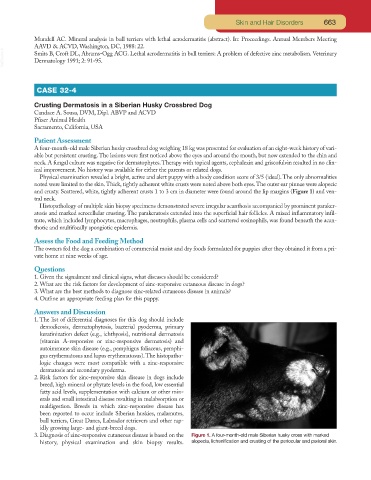
3. Diagnosis of zinc-responsive cutaneous disease is based on the Figure 1. A four-month-old male Siberian husky cross with marked
history, physical examination and skin biopsy results. alopecia, lichenification and crusting of the periocular and perioral skin.