
Page 645 - Small Animal Clinical Nutrition 5th Edition
P. 645
668 Small Animal Clinical Nutrition
commonly occurs in the shoulder (proximal humerus, Figure
VetBooks.ir 33-2), stifle (distal femur), hock (talus) and elbow (distal
humerus). Acute inflammatory joint disease (or degenerative
joint disease) may ensue subsequent to development of osteo-
chondrosis when the cartilage surface is disrupted and sub-
chondral bone is exposed to synovial fluid. Inflammatory
mediators and cartilage fragments are released into the joint
(osteochondritis dissecans), which perpetuates the cycle of
degenerative joint disease (Hill et al, 1984) (Chapter 34).
Other disease processes such as spondylolisthesis, intra-artic-
ular fracture, complete or partial epiphysiolysis and deformed
joint surfaces have been associated with osteochondrosis but
their etiology is still undetermined.
Elbow Dysplasia
Elbow dysplasia describes the four main developmental dys-
plastic conditions that are frequently diagnosed in the elbow
joint: 1) osteochondrosis of the medial condyle, 2) fragmented
medial coronoid process, 3) ununited anconeal process and 4)
elbow incongruency, due to a relatively short ulna. These con-
ditions result in severe lameness although the clinical manifes-
tation varies with the breed, the underlying diagnosis, the typ-
ical exercise pattern and the amount of osteoarthritis.
Breed specific distribution of the elbow dysplasia types has
been recognized and elbow dysplasia has a complex hereditary
background. Each of the conditions included may occur inde-
pendently in the canine population, although they can occur in
the same elbow in different combinations.
The frequency and severity of elbow dysplasia are also sub-
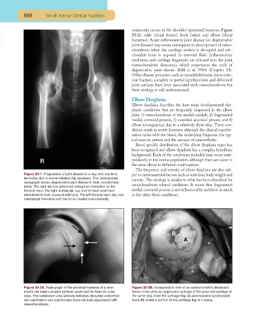
Figure 33-1. Progression of joint disease in a dog with rear-limb ject to environmental factors such as nutrition, body weight and
lameness due to severe bilateral hip dysplasia. This ventrodorsal exercise. The etiology is similar to what has been described for
radiograph shows degenerative joint disease in both coxofemoral
joints. The right hip has advanced osteophyte formation on the osteochondrosis-related conditions. It seems that fragmented
femoral neck. The right acetabular cup and femoral head have medial coronoid process is not influenced by nutrition as much
remodeled to form a pseudoarthrosis. The left femoral neck also has as the other three conditions.
osteophyte formation and the hip is luxated craniodorsally.
.
Figure 33-2A. Radiograph of the proximal humerus of a nine- Figure 33-2B. Intraoperative view of an osteochondritis dissecans
month-old male Labrador retriever examined for forelimb lame- lesion in the articular epiphyseal cartilage of the proximal cartilage of
ness. The radiolucent area (arrows) indicates disrupted endochon- the same dog. Note the cartilage flap (A) and exposed subchondral
dral ossification and subchondral bone necrosis associated with bone (B) where a portion of the cartilage flap is missing.
osteochondrosis.