
Page 17 - Essential Haematology
P. 17
Chapter 1 Haemopoiesis / 3
Pluripotent
stem cell
CFU GEMM
Common myeloid
progenitor cell
Common lymphoid
progenitor cell
CFU baso
BFU E
CFU GMEo
Erythroid CFU Eo
progenitors Eosinophil
progenitor
CFU GM
CFU Meg Granulocyte
Megakary- monocyte
CFU E Thymus
ocyte progenitor
progenitor
CFU-M CFU-G
B T NK
Red Platelets Mono- Neutro- Eosino- Baso- Lymphocytes NK cell
cells cytes phils phils phils
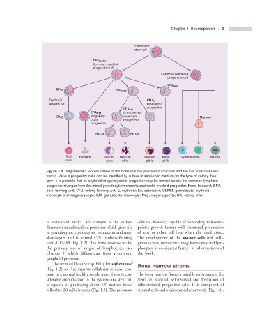
Figure 1.2 Diagrammatic representation of the bone marrow pluripotent stem cell and the cell lines that arise
from it. Various progenitor cells can be identifi ed by culture in semi - solid medium by the type of colony they
form. It is possible that an erythroid/megakaryocytic progenitor may be formed before the common lymphoid
progenitor diverges from the mixed granulocytic/monocyte/eosinophil myeloid progenitor. Baso, basophil; BFU,
burst - forming unit; CFU, colony - forming unit; E, erythroid; Eo, eosinophil; GEMM, granulocyte, erythroid,
monocyte and megakaryocyte; GM, granulocyte, monocyte; Meg, megakaryocyte; NK, natural killer.
in semi - solid media. An example is the earliest cells are, however, capable of responding to haemo-
detectable mixed myeloid precursor which gives rise poietic growth factors with increased production
to granulocytes, erythrocytes, monocytes and meg- of one or other cell line when the need arises.
akaryocytes and is termed CFU (colony - forming The development of the mature cells (red cells,
unit) - GEMM (Fig. 1.2 ). The bone marrow is also granulocytes, monocytes, megakaryocytes and lym-
the primary site of origin of lymphocytes (see phocytes) is considered further in other sections of
Chapter 9 ) which differentiate from a common this book.
lymphoid precursor.
The stem cell has the capability for self - renewal Bone m arrow s troma
(Fig. 1.3 ) so that marrow cellularity remains con-
stant in a normal healthy steady state. There is con- The bone marrow forms a suitable environment for
siderable amplification in the system: one stem cell stem cell survival, self - renewal and formation of
6
is capable of producing about 10 mature blood differentiated progenitor cells. It is composed of
cells after 20 cell divisions (Fig. 1.3 ). Th e precursor stromal cells and a microvascular network (Fig. 1.4 ).