
Page 429 - Basic _ Clinical Pharmacology ( PDFDrive )
P. 429
CHAPTER 24 Antiseizure Drugs 415
A1 A2
–10 mV
–80 mV
Closed
Fast inactivation (resting) Inactivated
Opening of of Na + channels
+
Na channels 0.5 nA
10 mV 2 ms
10 ms
Open (activated)
B
Closed (resting) Open (activated)
+ + + + Activation – – + + – –
Hyperpolarized + + + + + + + + + + Depolarized
– – – – Deactivation + + + +
Recovery Inactivation
– – + + – –
+ +
+ + Depolarized
+ + + +
Inactivated
C1
Outside
2 I II III IV 1
P-loop
NH 3 + NH 3 +
S1 S2 S3 S4S5 S6 S1 S2 S3 S4S5 S6 S1 S2 S3 S4S5 S6 S1 S2 S3 S4S5 S6
+ + + +
+ + + +
+ + + +
+ + + +
– OOC COO –
+ H N
3
COO –
Inside
C2
Outer pore
I P-loop
II IV
III
IV-S6
Selectivity
filter
III-S6
Lamotrigine
Inner pore
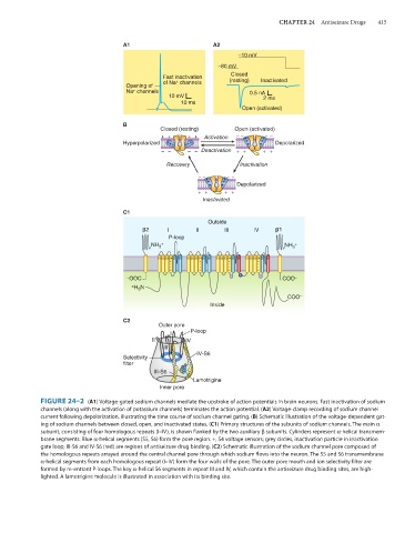
FIGURE 24–2 (A1) Voltage-gated sodium channels mediate the upstroke of action potentials in brain neurons. Fast inactivation of sodium
channels (along with the activation of potassium channels) terminates the action potential. (A2) Voltage-clamp recording of sodium channel
current following depolarization, illustrating the time course of sodium channel gating. (B) Schematic illustration of the voltage-dependent gat-
ing of sodium channels between closed, open, and inactivated states. (C1) Primary structures of the subunits of sodium channels. The main α
subunit, consisting of four homologous repeats (I–IV), is shown flanked by the two auxiliary β subunits. Cylinders represent α-helical transmem-
brane segments. Blue α-helical segments (S5, S6) form the pore region. +, S4 voltage sensors; grey circles, inactivation particle in inactivation
gate loop; III-S6 and IV-S6 (red) are regions of antiseizure drug binding. (C2) Schematic illustration of the sodium channel pore composed of
the homologous repeats arrayed around the central channel pore through which sodium flows into the neuron. The S5 and S6 transmembrane
α-helical segments from each homologous repeat (I–IV) form the four walls of the pore. The outer pore mouth and ion selectivity filter are
formed by re-entrant P-loops. The key α-helical S6 segments in repeat III and IV, which contain the antiseizure drug binding sites, are high-
lighted. A lamotrigine molecule is illustrated in association with its binding site.