
Page 590 - Basic _ Clinical Pharmacology ( PDFDrive )
P. 590
576 SECTION V Drugs That Act in the Central Nervous System
the ventral tegmental area (VTA), a tiny structure at the tip of the As a general rule, all addictive drugs activate the mesolimbic dopa-
brainstem, which projects to the nucleus accumbens, the amygdala, mine system. The behavioral significance of this increase of dopa-
the hippocampus, and the prefrontal cortex (Figure 32–1). Most mine is still debated. An appealing hypothesis is that mesolimbic
projection neurons of the VTA are dopamine-producing neurons. dopamine codes for the difference between expected and actual
When the dopamine neurons of the VTA begin to fire in bursts, reward and thus constitutes a strong learning signal (see Box: The
large quantities of dopamine are released in the nucleus accumbens Dopamine Hypothesis of Addiction).
and the prefrontal cortex. Early animal studies pairing electrical Since each addictive drug has a specific molecular target that
stimulation of the VTA with operant responses (eg, lever press- engages distinct cellular mechanisms to activate the mesolimbic
ing) that result in strong reinforcement established the central role system, three classes can be distinguished: A first group binds to
of the mesolimbic dopamine system in reward processing. Direct G protein-coupled receptors, a second group interacts with
io
application of drugs into the VTA also acts as a strong reinforcer, ionotropic receptors or ion channels, and a third group tar-
and systemic administration of drugs of abuse causes release of dopa- gets the dopamine transporter (Table 32–1 and Figure 32–2).
mine. Even selective activation of dopamine neurons is sufficient to G protein-coupled receptors (GPCRs) of the G family inhibit
io
drive reinforcement and elicit adaptive behavioral changes typically neurons through postsynaptic hyperpolarization and presynaptic
observed with addictive drugs. These very selective interventions regulation of transmitter release. These three classes of drugs
use optogenetic methods. Blue light is delivered in a freely mov- loosely map onto three distinct cellular mechanisms to increase
ing mouse through light guides to activate channelrhodopsin, a dopamine levels. The first is a direct stimulation of the dopamine
light-gated cation channel that is artificially expressed in dopamine neurons (eg, nicotine). The second mechanism is the interference
neurons. As a result, mice will self-administer light to activate VTA with the reuptake of dopamine or the promotion of nonvesicular
dopamine neurons. After several pairings with a specific environ- release (eg, amphetamines). This happens in the target regions
ment, a long-lasting place preference is established. Once the light is as well as the VTA itself, because dopamine neurons also express
no longer available, a seeking behavior is observed. Finally some mice somatodendritic transporters, which normally clear dopamine
will self-stimulate even if they have to endure a punishment (light released by the dendrites. Although drugs of this class also affect
electric shock). Conversely, using inhibitory optogenetic effectors or transporters of other monoamines (norepinephrine, serotonin),
activation of inhibitory neurons upstream causes aversion. action on the dopamine transporter remains central for addiction.
This is consistent with the observations that antidepressants that
block serotonin and norepinephrine uptake, but not dopamine
uptake, do not cause addiction even after prolonged use. The third
mechanism is indirect, whereby the drugs inhibit γ-aminobutyric
mPFC acid (GABA) neurons that act as local inhibitory interneurons
vHippo
(eg, opioids).
D1 LHb
DEPENDENCE: TOLERANCE &
D2 VP WITHDRAWAL
NAc RMTg
LDT With chronic exposure to addictive drugs, the brain shows signs
BLA VTA of adaptation. For example, if morphine is used at short intervals,
the dose has to be progressively increased over the course of several
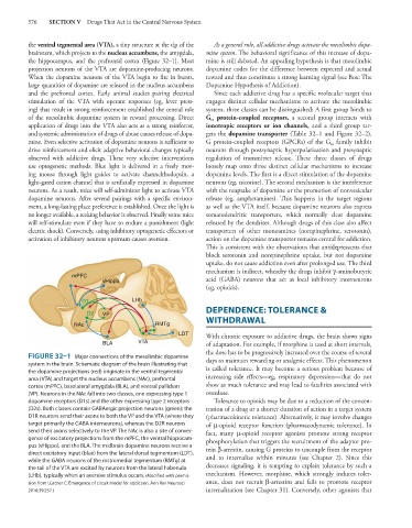
FIGURE 32–1 Major connections of the mesolimbic dopamine
system in the brain. Schematic diagram of the brain illustrating that days to maintain rewarding or analgesic effects. This phenomenon
the dopamine projections (red) originate in the ventral tegmental is called tolerance. It may become a serious problem because of
area (VTA) and target the nucleus accumbens (NAc), prefrontal increasing side effects—eg, respiratory depression—that do not
cortex (mPFC), basolateral amygdala (BLA), and ventral pallidum show as much tolerance and may lead to fatalities associated with
(VP). Neurons in the NAc fall into two classes, one expressing type 1 overdose.
dopamine receptors (D1s) and the other expressing type 2 receptors Tolerance to opioids may be due to a reduction of the concen-
(D2s). Both classes contain GABAergic projection neurons (green); the tration of a drug or a shorter duration of action in a target system
D1R neurons send their axons to both the VP and the VTA (where they (pharmacokinetic tolerance). Alternatively, it may involve changes
target primarily the GABA interneurons), whereas the D2R neurons of μ-opioid receptor function (pharmacodynamic tolerance). In
send their axons selectively to the VP. The NAc is also a site of conver- fact, many μ-opioid receptor agonists promote strong receptor
gence of excitatory projections from the mPFC, the ventral hippocam- phosphorylation that triggers the recruitment of the adaptor pro-
pus (vHippo), and the BLA. The midbrain dopamine neurons receive a
direct excitatory input (blue) from the lateral dorsal tegmentum (LDT), tein β-arrestin, causing G proteins to uncouple from the receptor
while the GABA neurons of the rostromedial tegmentum (RMTg) at and to internalize within minutes (see Chapter 2). Since this
the tail of the VTA are excited by neurons from the lateral habenula decreases signaling, it is tempting to explain tolerance by such a
(LHb), typically when an aversive stimulus occurs. (Modified with permis- mechanism. However, morphine, which strongly induces toler-
sion from Lüscher C: Emergence of circuit model for addiction. Ann Rev Neurosci ance, does not recruit β-arrestins and fails to promote receptor
2016;39:257.) internalization (see Chapter 31). Conversely, other agonists that