
Page 281 - parasitology for medical and clinical laboratoryprofessionals
P. 281
Laboratory Procedures for Identifying Parasitic Organisms and Their Ova 261
11. Perform a thorough examination for both preparations at low power, starting at
one corner of the cover slip and making sweeping moves either horizontally or
vertically in order to cover the entire slide. High-power objectives are used for
closer examinations of suspicious features.
12. Record the presence of white or red blood cells and any organisms observed on
the appropriate form. Any parasites observed should be reported by both genus
and species if possible and the stage of the parasite (ova, larvae, cyst, tropho-
zoite). Cellular components such as RBCs, WBCs, yeast, etc., are reported in a
semiquantitative form, as few, moderate, or many. Charcot-Leyden crystals are
also reported semiquantitatively.
Microscopic Examination of Wet Mount
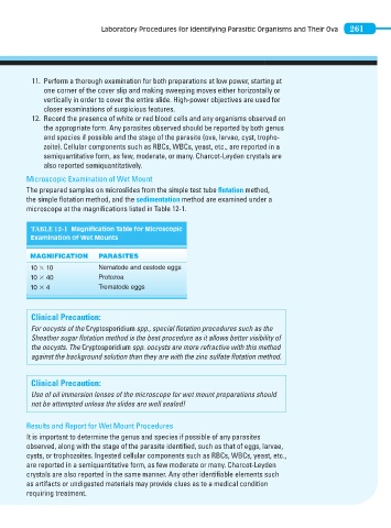
The prepared samples on microslides from the simple test tube fl otation method,
the simple flotation method, and the sedimentation method are examined under a
microscope at the magnifications listed in Table 12-1.
TABLE 12-1 Magnification Table for Microscopic
Examination of Wet Mounts
MAGNIFICATION PARASITES
10 3 10 Nematode and cestode eggs
10 3 40 Protozoa
10 3 4 Trematode eggs
Clinical Precaution:
For oocysts of the Cryptosporidium spp., special flotation procedures such as the
Sheather sugar flotation method is the best procedure as it allows better visibility of
the oocysts. The Cryptosporidium spp. oocysts are more refractive with this method
against the background solution than they are with the zinc sulfate fl otation method.
Clinical Precaution:
Use of oil immersion lenses of the microscope for wet mount preparations should
not be attempted unless the slides are well sealed!
Results and Report for Wet Mount Procedures
It is important to determine the genus and species if possible of any parasites
observed, along with the stage of the parasite identified, such as that of eggs, larvae,
cysts, or trophozoites. Ingested cellular components such as RBCs, WBCs, yeast, etc.,
are reported in a semiquantitative form, as few moderate or many. Charcot-Leyden
crystals are also reported in the same manner. Any other identifiable elements such
as artifacts or undigested materials may provide clues as to a medical condition
requiring treatment.