
Page 278 - parasitology for medical and clinical laboratoryprofessionals
P. 278
258 CHAPTER 12
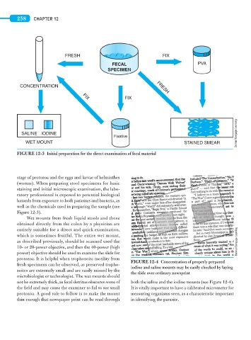
FRESH FIX
FECAL PVA
SPECIMEN
CONCENTRATION FRESH
FIX
FIX
Delmar/Cengage Learning
SALINE IODINE
Fixative
WET MOUNT STAINED SMEAR
FIGURE 12-3 Initial preparation for the direct examination of fecal material
stage of protozoa and the eggs and larvae of helminthes
(worms). When preparing stool specimens for basic
staining and initial microscopic examination, the labo-
ratory professional is exposed to potential biological
hazards from exposure to both parasites and bacteria, as
well as the chemicals used in preparing the sample (see
Figure 12-3).
Wet mounts from fresh liquid stools and those
obtained directly from the colon by a physician are
entirely suitable for a direct and quick examination,
which is sometimes fruitful. The entire wet mount,
as described previously, should be scanned used the Delmar/Cengage Learning
10- or 20-power objective, and then the 40-power (high
power) objective should be used to examine the slide for
protozoa. It is helpful when trophozoite motility from
FIGURE 12-4 Concentration of properly prepared
fresh specimens can be observed, as preserved tropho-
iodine and saline mounts may be easily checked by laying
zoites are extremely small and are easily missed by the
the slide over ordinary newsprint
microbiologist or technologist. The wet mounts should
not be extremely thick, as fecal detritus obscures some of both the saline and the iodine mounts (see Figure 12-4).
the field and may cause the examiner to fail to see small It is vitally important to have a calibrated micrometer for
protozoa. A good rule to follow is to make the mount measuring organisms seen, as a characteristic important
thin enough that newspaper print can be read through in identifying the parasite.