
Page 196 - Atlas of Histology with Functional Correlations
P. 196
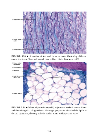
FIGURE  5.20  ■  A  section  of  the  wall  from  an  aorta  illustrating  different
               connective tissue fibers and smooth muscle fibers. Stain: blue stain. ×130.


               FIGURE 5.21 ■ White adipose tissue (cells) adjacent to skeletal muscle fibers
               and dense irregular collagen fibers. Histologic preparation dissolved the lipids in
               the cell cytoplasm, showing only the nuclei. Stain: Mallory-Azan. ×130.


                                                          195