
Page 234 - Atlas of Histology with Functional Correlations
P. 234
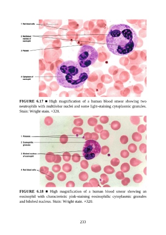
FIGURE  6.17  ■  High  magnification  of  a  human  blood  smear  showing  two
               neutrophils with multilobar nuclei and some light-staining cytoplasmic granules.
               Stain: Wright stain. ×320.


               FIGURE  6.18  ■  High  magnification  of  a  human  blood  smear  showing  an
               eosinophil  with  characteristic  pink-staining  eosinophilic  cytoplasmic  granules

               and bilobed nucleus. Stain: Wright stain. ×320.


                                                          233