
Page 262 - Atlas of Histology with Functional Correlations
P. 262
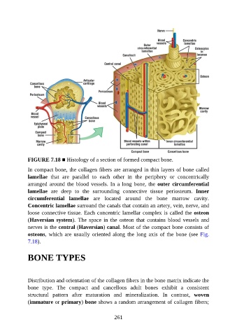
FIGURE 7.18 ■ Histology of a section of formed compact bone.

               In compact bone, the collagen fibers are arranged in thin layers of bone called

               lamellae  that  are  parallel  to  each  other  in  the  periphery  or  concentrically
               arranged around the blood vessels. In a long bone, the outer circumferential
               lamellae  are  deep  to  the  surrounding  connective  tissue  periosteum.  Inner

               circumferential  lamellae  are  located  around  the  bone  marrow  cavity.
               Concentric lamellae surround the canals that contain an artery, vein, nerve, and
               loose connective tissue. Each concentric lamellar complex is called the osteon
               (Haversian system).  The  space  in  the  osteon  that  contains  blood  vessels  and
               nerves is the central (Haversian) canal. Most of the compact bone consists of

               osteons,  which  are  usually  oriented  along  the  long  axis  of  the  bone  (see  Fig.
               7.18).


               BONE TYPES


               Distribution and orientation of the collagen fibers in the bone matrix indicate the
               bone  type.  The  compact  and  cancellous  adult  bones  exhibit  a  consistent

               structural  pattern  after  maturation  and  mineralization.  In  contrast,  woven
               (immature or primary) bone shows a random arrangement of collagen fibers;


                                                          261