
Page 331 - Atlas of Histology with Functional Correlations
P. 331
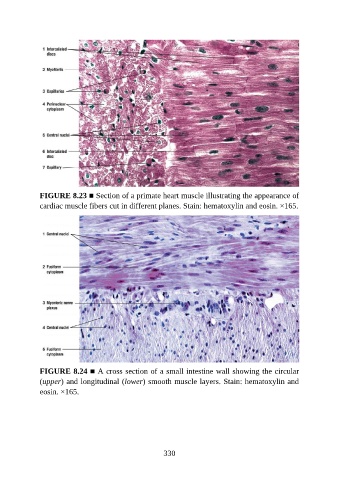
FIGURE 8.23 ■ Section of a primate heart muscle illustrating the appearance of
               cardiac muscle fibers cut in different planes. Stain: hematoxylin and eosin. ×165.


               FIGURE 8.24 ■ A cross section of a small intestine wall showing the circular
               (upper) and longitudinal (lower) smooth muscle layers. Stain: hematoxylin and
               eosin. ×165.


                                                          330