
Page 430 - Atlas of Histology with Functional Correlations
P. 430
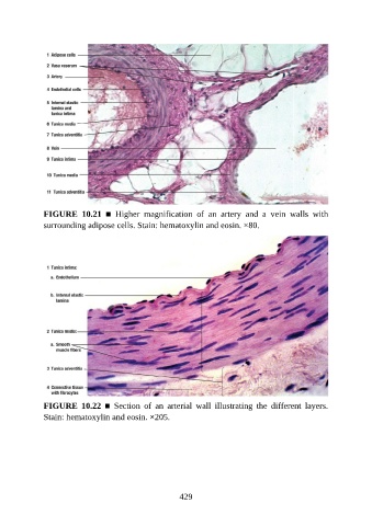
FIGURE  10.21  ■  Higher  magnification  of  an  artery  and  a  vein  walls  with
               surrounding adipose cells. Stain: hematoxylin and eosin. ×80.


               FIGURE  10.22  ■  Section  of  an  arterial  wall  illustrating  the  different  layers.

               Stain: hematoxylin and eosin. ×205.


                                                          429