
Page 436 - Atlas of Histology with Functional Correlations
P. 436
on the convex surface. Lymph is then filtered as it flows through the cortex and
medullary sinuses to exit the lymph node on the opposite side via the efferent
lymphatic vessels.
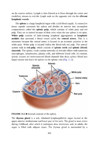
The spleen is a large lymphoid organ with a rich blood supply. A connective
tissue capsule surrounds the spleen and divides its interior into incomplete
compartments called the splenic pulp, which consists of white pulp and red
pulp. They are so named because of their color when the raw spleen is cut open.
White pulp consists of dark-staining lymphoid aggregations or lymphatic
nodules that surround a blood vessel called the central artery. This is a
misnomer because the central artery is located in an eccentric position in the
white pulp. White pulp is located within the blood-rich red pulp. The arterial
system ends in red pulp, which consists of splenic cords and splenic (blood)
sinusoids. The splenic cords contain networks of reticular fibers with numerous
macrophages, lymphocytes, plasma cells, and different blood cells. In contrast,
splenic sinuses are interconnected blood channels that drain splenic blood into
larger sinuses that leave the spleen via the splenic vein (Fig. 11.3).
FIGURE 11.3 ■ Internal contents of the spleen.
The thymus gland is a soft, lobulated lymphoepithelial organ located in the
upper anterior mediastinum and lower part of the neck. The gland is most active
during childhood, after which it undergoes slow involution, and, in adults, the
organ is filled with adipose tissue. The thymus gland is surrounded by a
435