
Page 441 - Atlas of Histology with Functional Correlations
P. 441
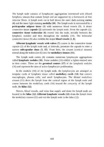
The  lymph  node  consists  of  lymphocyte  aggregations  intermixed  with  dilated

               lymphatic sinuses that contain lymph and are supported by a framework of fine
               reticular fibers. A lymph node cut in half shows the outer dark-staining cortex
               (4) and the inner light-staining medulla (10). The lymph node is surrounded by a
               pericapsular  adipose  tissue  (1)  with  numerous  blood  vessels  (9).  A  dense
               connective tissue capsule (2) surrounds the lymph node. From the capsule (2),

               connective  tissue  trabeculae  (6)  extend  into  the  node,  initially  between  the
               lymphatic  nodules  and  then  throughout  the  medulla  (10).  The  trabecular
               connective tissue (6) also exhibits the major blood vessels (5, 8).

                   Afferent lymphatic vessels with valves (7) course in the connective tissue

               capsule (2) of the lymph node and, at intervals, penetrate the capsule to enter a
               narrow  subcapsular  sinus  (3,  15).  From  here,  the  sinuses  (cortical  sinuses)
               extend along the trabeculae (6) into the medullary sinuses (11).

                   The  lymph  node  cortex  (4)  contains  numerous  lymphocyte  aggregations
               called lymphatic nodules (16). Some nodules (16) exhibit a lighter-stained area

               in their center. These are the germinal centers (17) of the lymphatic nodules
               (16) and represent the sites of active lymphocyte proliferation.

                   In  the  medulla  (10)  of  the  lymph  node,  the  lymphocytes  are  arranged  as
               irregular  cords  of  lymphatic  tissue  called  medullary  cords  (14)  that  contain

               macrophages,  plasma  cells,  and  small  lymphocytes.  The  dilated  medullary
               sinuses  (11)  drain  the  lymph  from  the  cortical  region  of  the  lymph  node  and
               course  between  the  medullary  cords  (14)  toward  the  concavity  of  the  lymph
               node, the hilus (12).

                   Nerves, blood vessels, and veins that supply and drain the lymph node are

               located in the hilus (12). Efferent lymphatic vessels (13) drain the lymph from
               the medullary sinuses (11) and exit the lymph node in the hilus (12).


                                                          440