
Page 446 - Atlas of Histology with Functional Correlations
P. 446
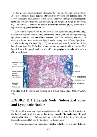
This low-power photomicrograph illustrates the lymph node cortex and medulla.

               A loose connective tissue capsule (4) with blood vessels and adipose cells (7)
               covers the lymph node. Inferior to the capsule (4) is the subcapsular (marginal)
               sinus (5), which overlies the darker-staining and peripheral lymph node cortex
               (3). The cortex (3) exhibits numerous lymphatic nodules (1, 6), some with a
               lighter-staining germinal center (2).


                   The  central  region  of  the  lymph  node  is  the  lighter-staining  medulla  (9),
               characterized by the dark-staining medullary cords (12) and the light-staining
               lymphatic  channels,  the  medullary  sinuses  (11).  The  medullary  sinuses  (11)
               drain  the  lymph  that  enters  the  lymph  node  through  the  afferent  lymphatic

               vessels  in  the  capsule  (see  Fig.  11.5)  and  converges  toward  the  hilum  of  the
               lymph node (see Fig. 11.2) that contains numerous arteries (8) and veins. The
               lymph  leaves  the  lymph  node  via  the  efferent  lymphatic  vessels  with  valves
               (10) at the hilum.


               FIGURE  11.6  ■  Cortex  and  medulla  of  a  lymph  node.  Stain:  Mallory-Azan.
               ×25.


               FIGURE  11.7  |  Lymph  Node:  Subcortical  Sinus


               and Lymphatic Nodule


               This figure illustrates, at a higher magnification and in greater detail, a portion of
               the  lymph  node  with  the  connective  tissue  capsule  (3),  trabecula  (4),  and
               subcapsular  sinus  (1)  that  continue  on  both  sides  of  the  trabecula  (4)  as

               trabecular sinuses (12) into the interior of the lymph node.

                   The reticular connective tissue, the reticular cells (8, 11), is seen in different


                                                          445