
Page 451 - Atlas of Histology with Functional Correlations
P. 451
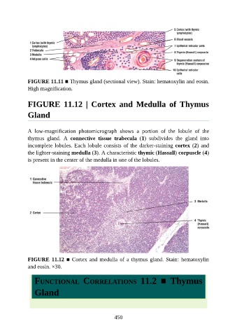
FIGURE 11.11 ■ Thymus gland (sectional view). Stain: hematoxylin and eosin.
               High magnification.


               FIGURE  11.12  |  Cortex  and  Medulla  of  Thymus

               Gland


               A  low-magnification  photomicrograph  shows  a  portion  of  the  lobule  of  the

               thymus  gland.  A  connective  tissue  trabecula  (1)  subdivides  the  gland  into
               incomplete lobules. Each lobule consists of the darker-staining cortex (2) and
               the lighter-staining medulla (3). A characteristic thymic (Hassall) corpuscle (4)
               is present in the center of the medulla in one of the lobules.


               FIGURE 11.12 ■ Cortex and medulla of a thymus gland. Stain: hematoxylin
               and eosin. ×30.


                  FUNCTIONAL  CORRELATIONS  11.2  ■  Thymus


                  Gland


                                                          450