Page 455 - Atlas of Histology with Functional Correlations
P. 455
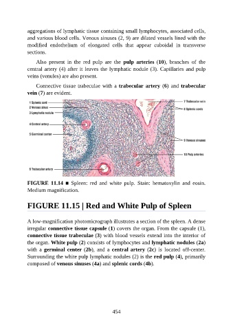
aggregations of lymphatic tissue containing small lymphocytes, associated cells,

               and various blood cells. Venous sinuses (2, 9) are dilated vessels lined with the
               modified  endothelium  of  elongated  cells  that  appear  cuboidal  in  transverse
               sections.

                   Also  present  in  the  red  pulp  are  the  pulp  arteries  (10),  branches  of  the
               central artery (4) after it leaves the lymphatic nodule (3). Capillaries and pulp

               veins (venules) are also present.

                   Connective tissue trabeculae with a trabecular artery (6) and trabecular
               vein (7) are evident.


               FIGURE  11.14  ■  Spleen:  red  and  white  pulp.  Stain:  hematoxylin  and  eosin.
               Medium magnification.


               FIGURE 11.15 | Red and White Pulp of Spleen


               A low-magnification photomicrograph illustrates a section of the spleen. A dense
               irregular connective tissue capsule (1) covers the organ. From the capsule (1),
               connective tissue trabeculae (3) with blood vessels extend into the interior of

               the organ. White pulp (2) consists of lymphocytes and lymphatic nodules (2a)
               with  a  germinal center (2b),  and  a  central artery (2c)  is  located  off-center.
               Surrounding the white pulp lymphatic nodules (2) is the red pulp (4), primarily
               composed of venous sinuses (4a) and splenic cords (4b).


                                                          454

