
Page 450 - Atlas of Histology with Functional Correlations
P. 450
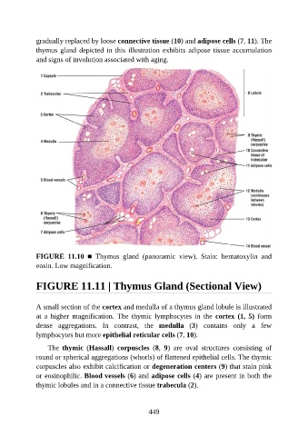
gradually replaced by loose connective tissue (10) and adipose cells (7, 11). The

               thymus gland depicted in this illustration exhibits adipose tissue accumulation
               and signs of involution associated with aging.


               FIGURE  11.10  ■  Thymus  gland  (panoramic  view).  Stain:  hematoxylin  and

               eosin. Low magnification.


               FIGURE 11.11 | Thymus Gland (Sectional View)


               A small section of the cortex and medulla of a thymus gland lobule is illustrated

               at  a  higher  magnification.  The  thymic  lymphocytes  in  the  cortex  (1,  5)  form
               dense  aggregations.  In  contrast,  the  medulla  (3)  contains  only  a  few
               lymphocytes but more epithelial reticular cells (7, 10).

                   The  thymic  (Hassall)  corpuscles  (8,  9)  are  oval  structures  consisting  of

               round or spherical aggregations (whorls) of flattened epithelial cells. The thymic
               corpuscles also exhibit calcification or degeneration centers (9) that stain pink
               or eosinophilic. Blood vessels (6) and adipose cells (4) are present in both the
               thymic lobules and in a connective tissue trabecula (2).


                                                          449