
Page 449 - Atlas of Histology with Functional Correlations
P. 449
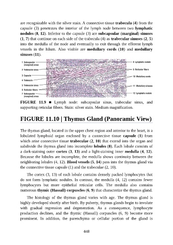
are recognizable with the silver stain. A connective tissue trabecula (4) from the

               capsule  (3)  penetrates  the  interior  of  the  lymph  node  between  two  lymphatic
               nodules (8, 12). Inferior to the capsule (3) are subcapsular (marginal) sinuses
               (1, 7) that continue on each side of the trabecula (4) as trabecular sinuses (2, 5)
               into the medulla of the node and eventually to exit through the efferent lymph
               vessels  in  the  hilum.  Also  visible  are  medullary  cords  (10)  and  medullary

               sinuses (11).


               FIGURE  11.9  ■  Lymph  node:  subcapsular  sinus,  trabecular  sinus,  and

               supporting reticular fibers. Stain: silver stain. Medium magnification.


               FIGURE 11.10 | Thymus Gland (Panoramic View)


               The thymus gland, located in the upper chest region and anterior to the heart, is a

               lobulated  lymphoid  organ  enclosed  by  a  connective  tissue  capsule  (1)  from
               which arise connective tissue trabeculae (2, 10) that extend into the organ and
               subdivide the thymus gland into incomplete lobules (8). Each lobule consists of
               a dark-staining outer cortex (3, 13) and a light-staining inner medulla (4, 12).

               Because the lobules are incomplete, the medulla shows continuity between the
               neighboring lobules (4, 12). Blood vessels (5, 14) pass into the thymus gland via
               the connective tissue capsule (1) and the trabeculae (2, 10).

                   The cortex (3, 13) of each lobule contains densely packed lymphocytes that
               do not form lymphatic nodules. In contrast, the medulla (4, 12) contains fewer

               lymphocytes  but  more  epithelial  reticular  cells.  The  medulla  also  contains
               numerous thymic (Hassall) corpuscles (6, 9) that characterize the thymus gland.

                   The  histology  of  the  thymus  gland  varies  with  age.  The  thymus  gland  is
               highly developed shortly after birth. By puberty, thymus glands begin to involute

               with  gradual  regression  and  degeneration.  As  a  consequence,  lymphocyte
               production  declines,  and  the  thymic  (Hassall)  corpuscles  (6,  9)  become  more
               prominent.  In  addition,  the  parenchyma  or  cellular  portion  of  the  gland  is


                                                          448