
Page 453 - Atlas of Histology with Functional Correlations
P. 453
gland, which are characteristic features in identifying the thymus gland. It is
believed that thymic corpuscles produce cytokine thymic stromal
lymphopoietin that induces antigen-presenting cells ([APCs] also known as
dendritic cells) to promote development of the regulatory T cells. The thymus
gland involutes after puberty and becomes filled with adipose tissue, and the
production of T cells decreases. However, because T-lymphocyte progeny
has been established, immunity is maintained without new T-cell production.
However, if the thymus gland is removed from a newborn, the lymphoid
organs will not receive the immunocompetent T cells, and the individual will
not acquire the immunologic competence to fight pathogens. Death may
occur early in life as a result of complications of an infection and the lack of
a functional immune system.
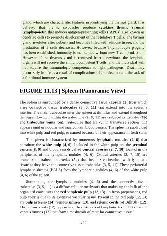
FIGURE 11.13 | Spleen (Panoramic View)
The spleen is surrounded by a dense connective tissue capsule (1) from which
arise connective tissue trabeculae (3, 5, 11) that extend into the spleen’s
interior. The main trabeculae enter the spleen at the hilus and extend throughout
the organ. Located within the trabeculae (3, 5, 11) are trabecular arteries (5b)
and trabecular veins (5a). Trabeculae that are cut in transverse section (11)
appear round or nodular and may contain blood vessels. The spleen is subdivided
into white pulp and red pulp, so named because of their appearance in fresh state.
The spleen is characterized by numerous lymphatic nodules (4, 6) that
constitute the white pulp (4, 6). Included in the white pulp are the germinal
centers (8, 9) and blood vessels called central arteries (2, 7, 10) located in the
peripheries of the lymphatic nodules (4, 6). Central arteries (2, 7, 10) are
branches of trabecular arteries (5b) that become ensheathed with lymphatic
tissue as they leave the connective tissue trabeculae (3, 5, 11). These periarterial
lymphatic sheaths (PALS) form the lymphatic nodules (4, 6) of the white pulp
(4, 6) of the spleen.
Surrounding the lymphatic nodules (4, 6) and the connective tissue
trabeculae (3, 5, 11) is a diffuse cellular meshwork that makes up the bulk of the
organ and constitutes the red or splenic pulp (12, 13). In fresh preparations, red
pulp color is due to its extensive vascular tissue. Present in the red pulp (12, 13)
are pulp arteries (14), venous sinuses (13), and splenic cords (of Billroth) (12).
The splenic cords (12) appear as diffuse strands of lymphatic tissue between the
venous sinuses (13) that form a meshwork of reticular connective tissue.
452