
Page 454 - Atlas of Histology with Functional Correlations
P. 454
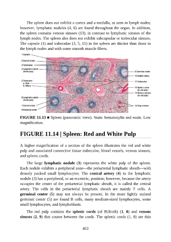
The spleen does not exhibit a cortex and a medulla, as seen in lymph nodes;

               however, lymphatic nodules (4, 6) are found throughout the organ. In addition,
               the spleen contains venous sinuses (13), in contrast to lymphatic sinuses of the
               lymph nodes. The spleen also does not exhibit subcapsular or trabecular sinuses.
               The capsule (1) and trabeculae (3, 5, 11) in the spleen are thicker than those in
               the lymph nodes and with some smooth muscle fibers.


               FIGURE 11.13 ■ Spleen (panoramic view). Stain: hematoxylin and eosin. Low

               magnification.


               FIGURE 11.14 | Spleen: Red and White Pulp


               A higher magnification of a section of the spleen illustrates the red and white
               pulp and associated connective tissue trabeculae, blood vessels, venous sinuses,
               and splenic cords.

                   The  large  lymphatic  nodule  (3)  represents  the  white  pulp  of  the  spleen.
               Each nodule exhibits a peripheral zone—the periarterial lymphatic sheath—with

               densely  packed  small  lymphocytes.  The  central  artery  (4)  in  the  lymphatic
               nodule (3) has a peripheral, or an eccentric, position; however, because the artery
               occupies the center of the periarterial lymphatic sheath, it is called the central

               artery.  The  cells  in  the  periarterial  lymphatic  sheath  are  mainly  T  cells.  A
               germinal  center  (5)  may  not  always  be  present.  In  the  more  lightly  stained
               germinal center (5) are found B cells, many medium-sized lymphocytes, some
               small lymphocytes, and lymphoblasts.

                   The  red  pulp  contains  the  splenic  cords  (of  Billroth)  (1,  8)  and  venous

               sinuses (2, 9) that course between the cords. The splenic cords (1, 8) are thin


                                                          453