
Page 456 - Atlas of Histology with Functional Correlations
P. 456
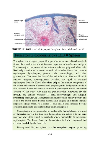
FIGURE 11.15 ■ Red and white pulp of the spleen. Stain: Mallory-Azan. ×21.


                  FUNCTIONAL CORRELATIONS 11.3 ■ Spleen


                 The spleen is the largest lymphoid organ with an extensive blood supply. It
                 filters  blood  and  is  the  site  of  immune  responses  to  blood-borne  antigens.
                 The two major components of the spleen are the red pulp and white pulp.

                 Red  pulp  consists  of  a  dense  network  of  reticular  fibers  that  contains
                 erythrocytes,  lymphocytes,  plasma  cells,  macrophages,  and  other
                 granulocytes.  The  main  function  of  the  red  pulp  is  to  filter  the  blood.  It
                 removes  antigens,  microorganisms,  platelets,  and  aged  or  abnormal
                 erythrocytes from the blood. The white pulp is the immune component of

                 the spleen and consists of accumulated lymphocytes in the lymphatic nodules
                 that surround the central artery or arteriole. Lymphocytes around the central
                 arteries  of  the  white  pulp  form  the  periarteriolar  lymphatic  sheaths

                 (PALS)  and  contain  primarily  T  cells,  macrophages,  and  antigen-
                 presenting cells (APCs). The lymphatic nodules contain mainly B cells. The
                 cells in the spleen detect trapped bacteria and antigens and initiate immune
                 responses  against  them.  As  a  result,  T  cells  and  B  cells  interact,  become
                 activated, proliferate, and perform their immune response.


                     Macrophages in the spleen also break down the hemoglobin of worn-out
                 erythrocytes, recycle the iron from hemoglobin, and return it to the bone
                 marrow, where it is reused for synthesis of new hemoglobin by developing
                 erythrocytes.  The  heme  from  the  hemoglobin  is  further  degraded  and

                 excreted into bile by the liver cells.

                     During  fetal  life,  the  spleen  is  a  hematopoietic  organ,  producing


                                                          455