
Page 448 - Atlas of Histology with Functional Correlations
P. 448
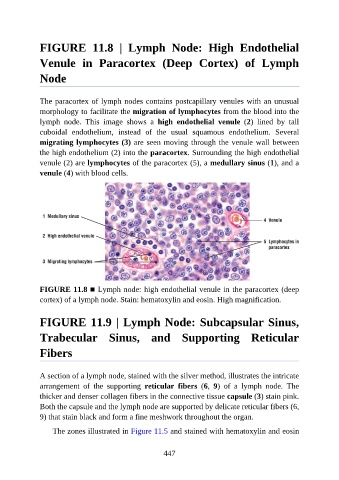
FIGURE  11.8  |  Lymph  Node:  High  Endothelial

               Venule  in  Paracortex  (Deep  Cortex)  of  Lymph

               Node


               The paracortex of lymph nodes contains postcapillary venules with an unusual
               morphology to facilitate the migration of lymphocytes from the blood into the
               lymph  node.  This  image  shows  a  high  endothelial  venule  (2)  lined  by  tall
               cuboidal  endothelium,  instead  of  the  usual  squamous  endothelium.  Several
               migrating lymphocytes (3) are seen moving through the venule wall between

               the high endothelium (2) into the paracortex. Surrounding the high endothelial
               venule (2) are lymphocytes of the paracortex (5), a medullary sinus (1), and a
               venule (4) with blood cells.


               FIGURE 11.8 ■ Lymph node: high endothelial venule in the paracortex (deep
               cortex) of a lymph node. Stain: hematoxylin and eosin. High magnification.


               FIGURE 11.9 | Lymph Node: Subcapsular Sinus,

               Trabecular  Sinus,  and  Supporting  Reticular

               Fibers


               A section of a lymph node, stained with the silver method, illustrates the intricate
               arrangement  of  the  supporting  reticular  fibers  (6,  9)  of  a  lymph  node.  The
               thicker and denser collagen fibers in the connective tissue capsule (3) stain pink.
               Both the capsule and the lymph node are supported by delicate reticular fibers (6,

               9) that stain black and form a fine meshwork throughout the organ.

                   The zones illustrated in Figure 11.5 and stained with hematoxylin and eosin


                                                          447