
Page 447 - Atlas of Histology with Functional Correlations
P. 447
regions of the node. Reticular cells (8, 11) are visible in the subcapsular sinus
(1), trabecular sinuses (12), and the germinal center (9) of the lymphatic
nodule (14). Numerous free macrophages (2, 6, 16) are also seen in the
subcapsular sinus (1), trabecular sinuses (12), and the germinal center (9) of the
lymphatic nodule (14).
A lymphatic nodule with a small section of its peripheral zone (14) and a
germinal center (9) with developing lymphocytes are visible. Endothelial cells
(5, 13) line the sinuses (1, 12) and form an incomplete cover over the surface of
the lymphatic nodules (14).
The dense peripheral zone of the lymphatic nodule (14) contains an
aggregation of small lymphocytes (7), characterized by dark-staining nuclei,
condensed chromatin, and little or no cytoplasm. Small lymphocytes (7) are also
present in the subcapsular sinus (1) and trabecular sinuses (12).
The germinal center (9) of the lymphatic nodule (14) contains medium-sized
lymphocytes (10) characterized by larger, lighter nuclei and more cytoplasm
than in the small lymphocytes (7). The nuclei of medium-sized lymphocytes (10)
exhibit variations in the size and density of the chromatin. The largest cells, with
less condensed chromatin, are the lymphoblasts (17) visible in the germinal
center (9) as large cells with a broad band of cytoplasm and a large vesicular
nucleus with one or more nucleoli. Lymphoblasts (15) produce other
lymphoblasts and medium-sized lymphocytes (10). With mitotic divisions of
lymphoblasts (15), the chromatin condenses and the cells decrease in size,
producing small lymphocytes (7).
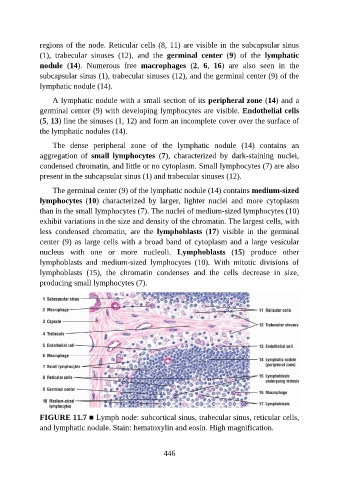
FIGURE 11.7 ■ Lymph node: subcortical sinus, trabecular sinus, reticular cells,
and lymphatic nodule. Stain: hematoxylin and eosin. High magnification.
446