
Page 442 - Atlas of Histology with Functional Correlations
P. 442
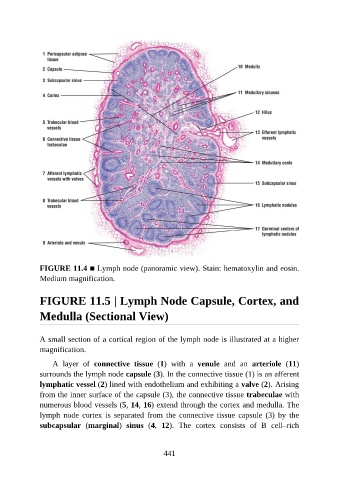
FIGURE 11.4 ■ Lymph node (panoramic view). Stain: hematoxylin and eosin.
               Medium magnification.


               FIGURE 11.5 | Lymph Node Capsule, Cortex, and

               Medulla (Sectional View)


               A small section of a cortical region of the lymph node is illustrated at a higher
               magnification.

                   A  layer  of  connective  tissue  (1)  with  a  venule  and  an  arteriole  (11)
               surrounds the lymph node capsule (3). In the connective tissue (1) is an afferent

               lymphatic vessel (2) lined with endothelium and exhibiting a valve (2). Arising
               from the inner surface of the capsule (3), the connective tissue trabeculae with
               numerous blood vessels (5, 14, 16) extend through the cortex and medulla. The
               lymph  node  cortex  is  separated  from  the  connective  tissue  capsule  (3)  by  the

               subcapsular  (marginal)  sinus  (4,  12).  The  cortex  consists  of  B  cell–rich


                                                          441